Finlay's Case Presentation

Welcome to my January 2020 Newsletter Case Presentation

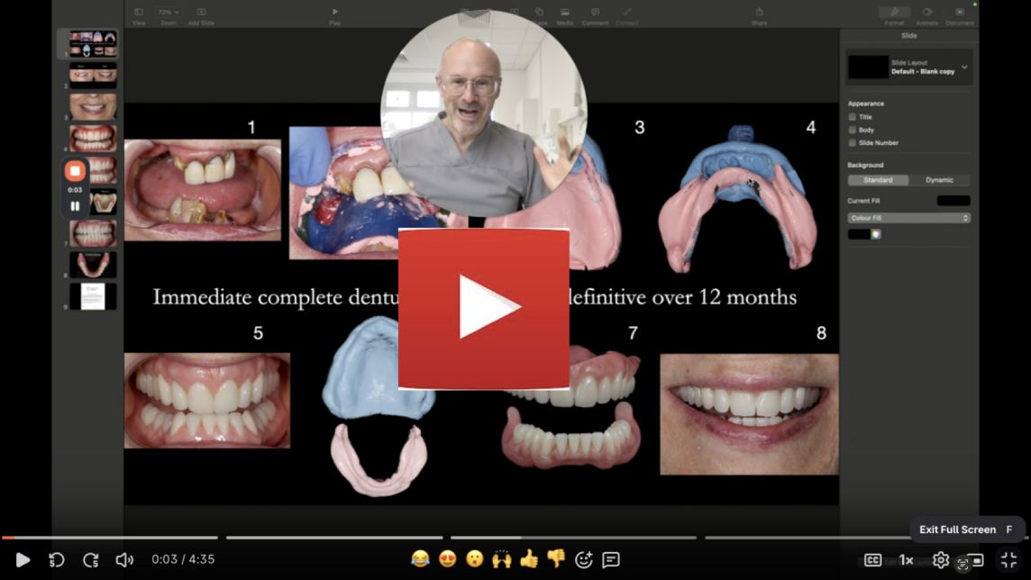
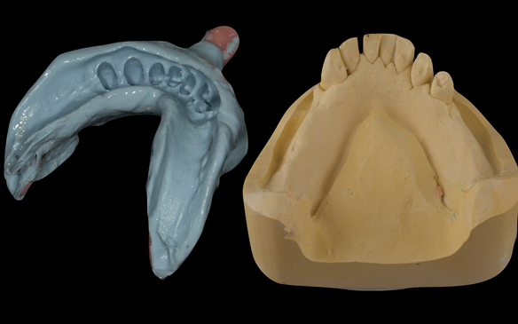
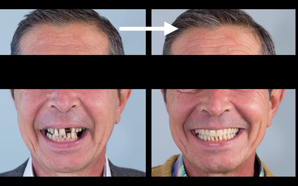
This newsletter describes in step by step detail the transition from acrylic based immediate dentures to metal based definitive dentures.

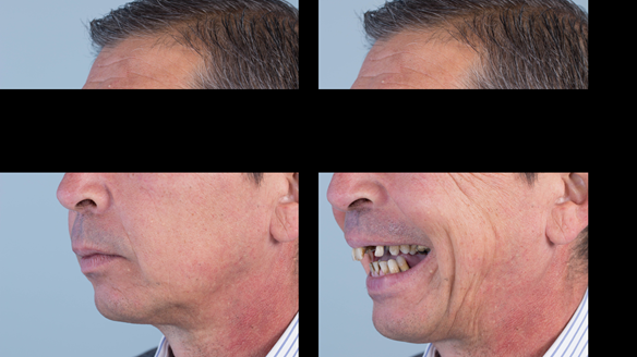
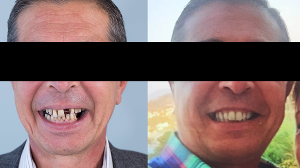
This 52 year old man was referred to me from his general dental practitioner in 2018.

Dental History and Concerns

10 years prior to consultation the patient's general dentist diagnosed periodontitis and referred him to Manchester Dental Hospital. He received a treatment plan to manage the periodontitis. Unfortunately, this was not acted on. Four years ago gaps developed between the upper front teeth. Orthodontic treatment was provided to align the teeth. A bonded retainer was fitted onto the upper front teeth. Approximately one year prior to consultation with me the patient noticed the teeth moving again. He consulted his orthodontist, who advised no further orthodontic treatment. One month prior to the consultation with me the upper left central incisor fell out whilst eating.

Social History

Cigarette smoker. 20 per day continuously for over 30 years

Dental wish list

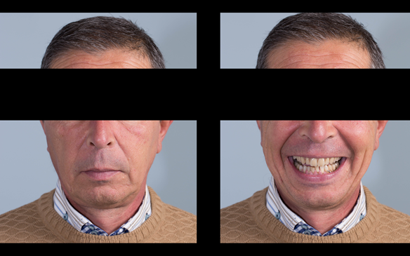
  1. "Something fixed which I don’t have to worry about"
  2. "To be able to smile and be confident"
  3. "I want teeth to look totally natural and healthy"
  4. "I don’t want to have a cosmetic look – but to look just how they were 2 years ago."

Diagnoses

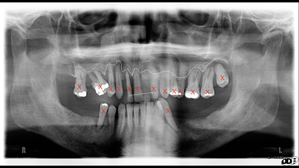
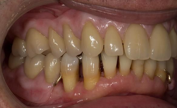
  1. Generalised periodontitis; stage IV grade C: currently unstable, risk factors: smoker.
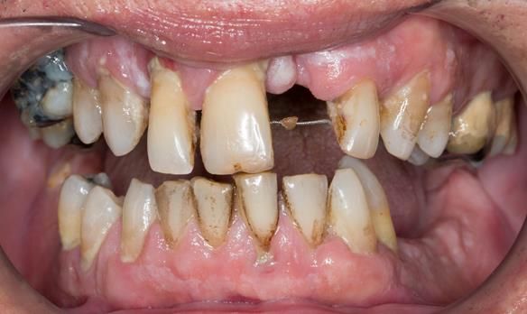
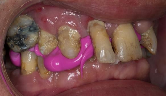
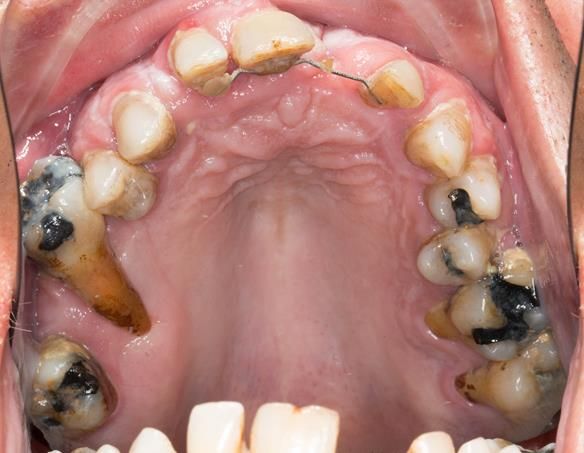
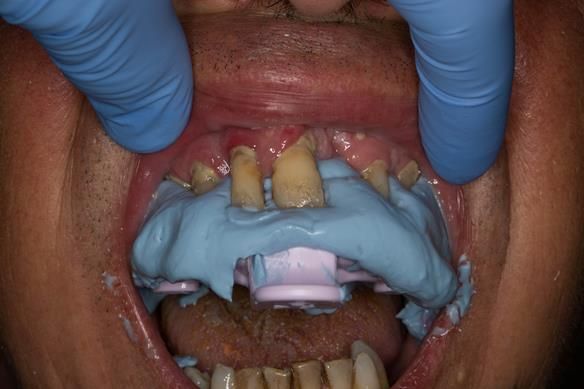
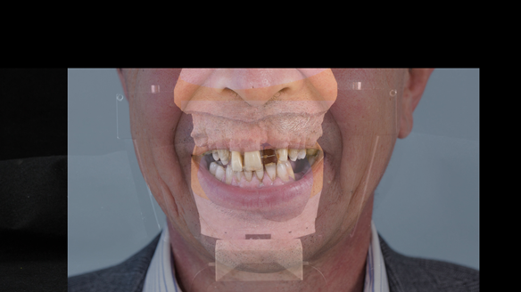
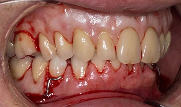
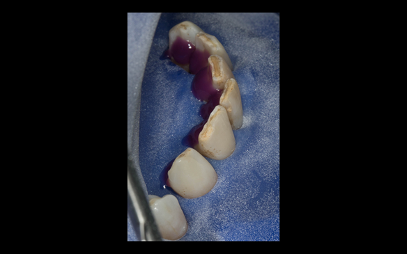
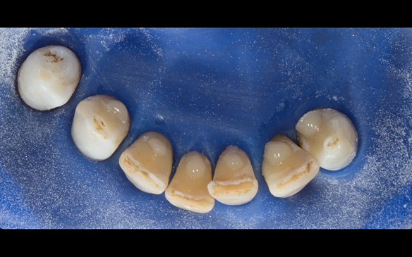
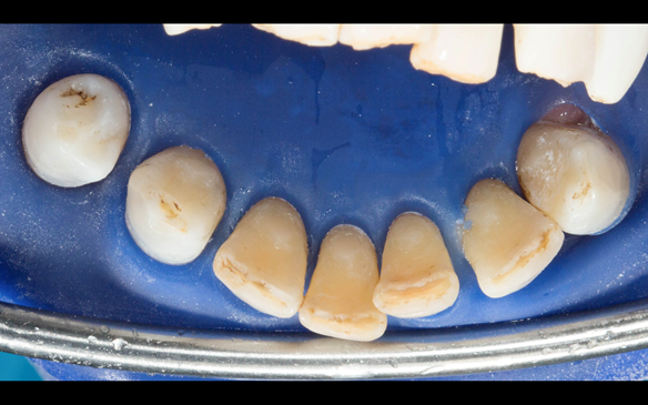
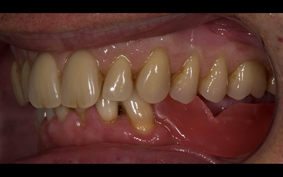
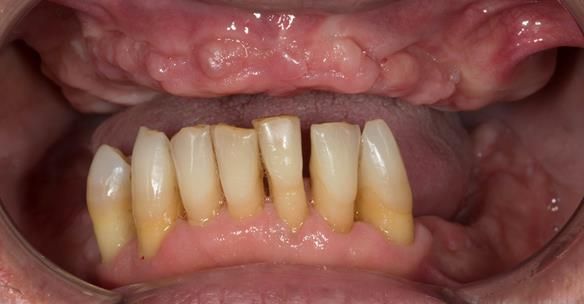
  2. The remaining maxillary teeth had hopeless prognosis in the short term. They exhibited 80 - 100% alveolar bone loss with increased mobility (Grade 2 - 3).
  3. The lower right second premolar and lower left first premolar (LR5 LL4) had hopeless prognosis in the short term. They exhibited 80 - 100% alveolar bone loss with grade 3 mobility.
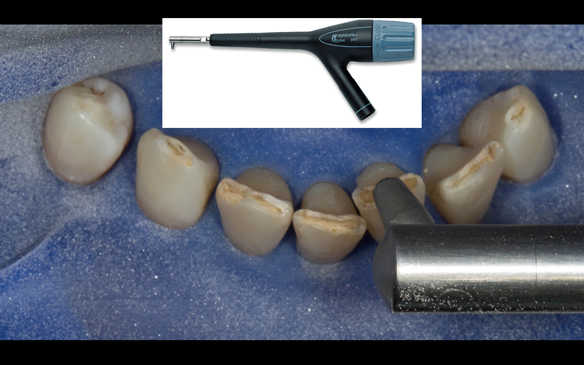
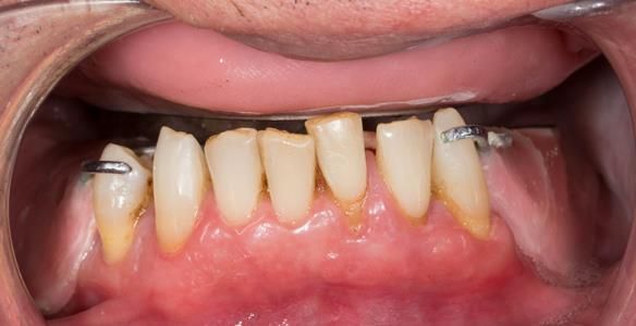
  4. The remaining mandibular teeth had approximately 30 - 50% alveolar bone loss with grade 1 mobility.

I did not believe that dental implant supported restorations (fixed or removable) were in this patients best interest given the history and extent of the periodontal disease and the smoking history. I advised him that other practitioners may be willing to provide implant supported fixed/removable restorations as an option. He was given time and space to choose his preferred option for valid consent.

Following two discussion appointments with me, the patient decided to have the following treatment plan:
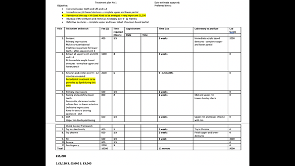
Treatment plan

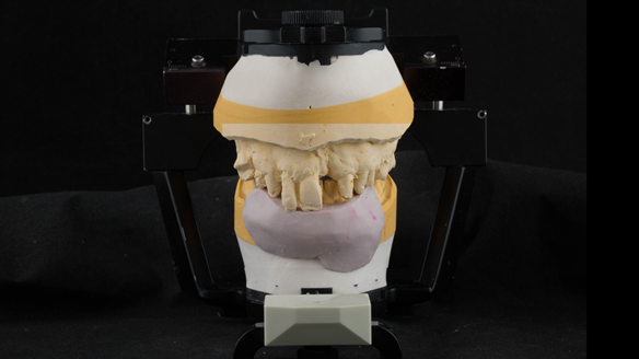
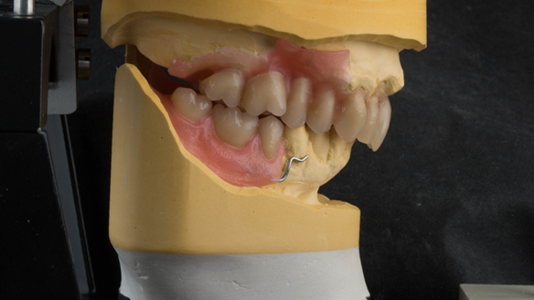
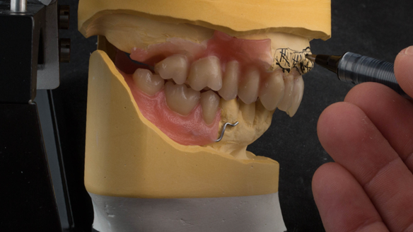
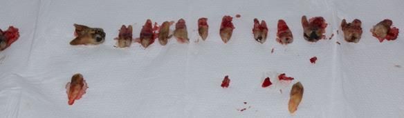
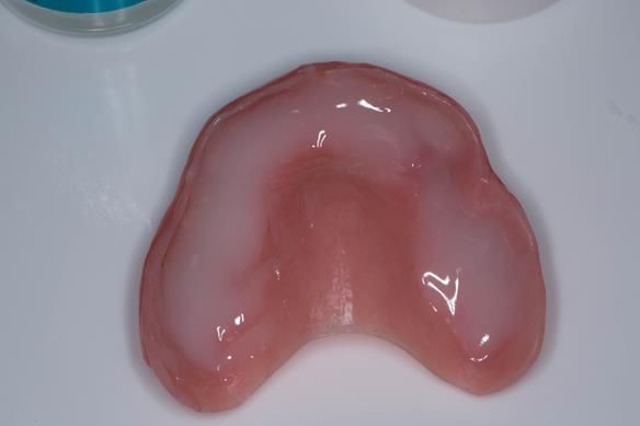
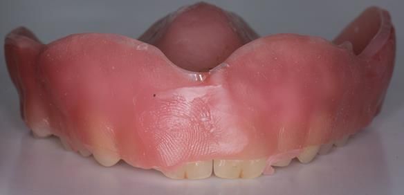
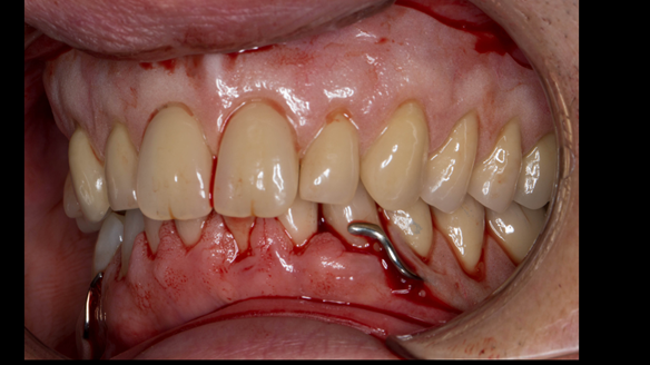
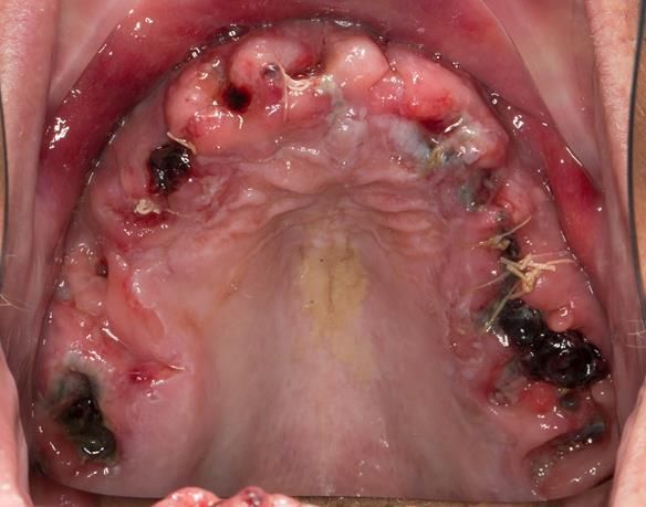
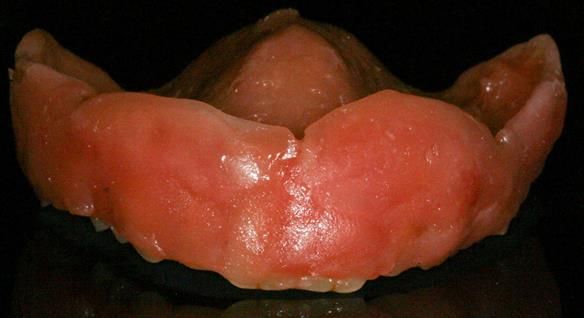
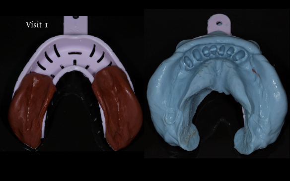
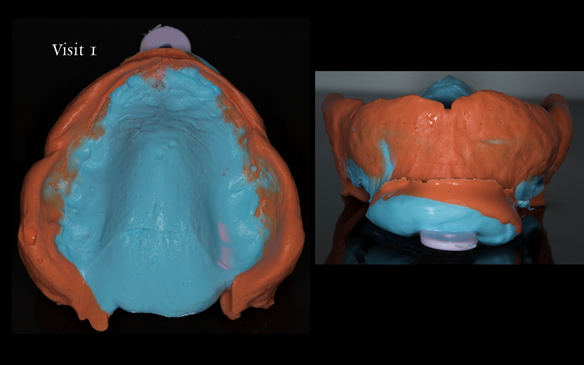
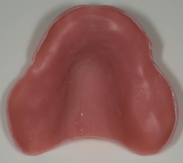
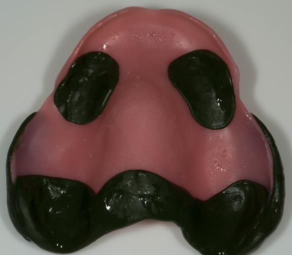
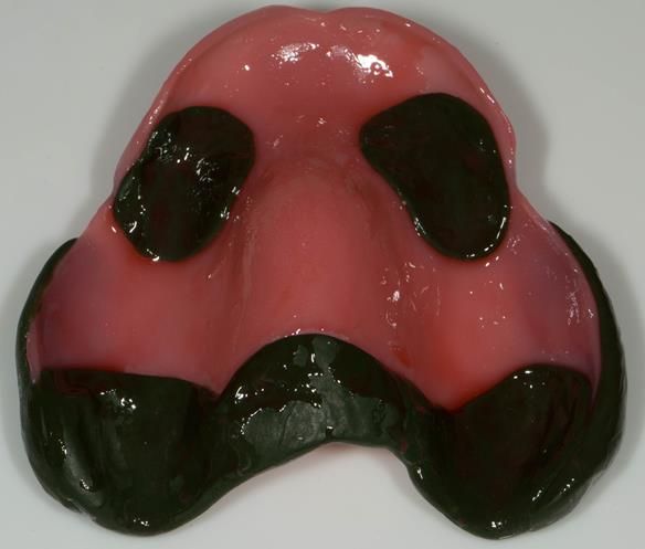
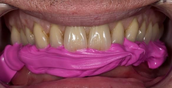
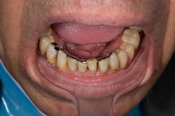
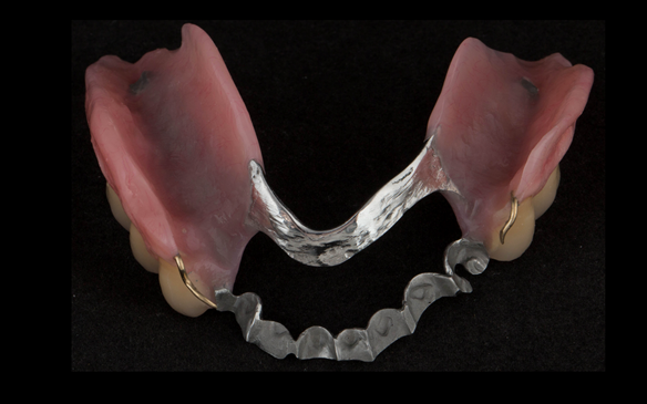
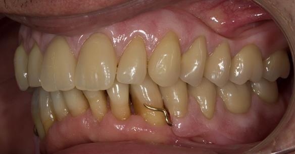
  1. Extraction of all upper teeth and LR5 and LL4 and fitting of immediate acrylic based (Mk 1) dentures - complete upper and lower partial
  2. Periodontal therapy involving smoking cessation advice – with Mr Syed Abad, Specialist in Periodontics at the practice
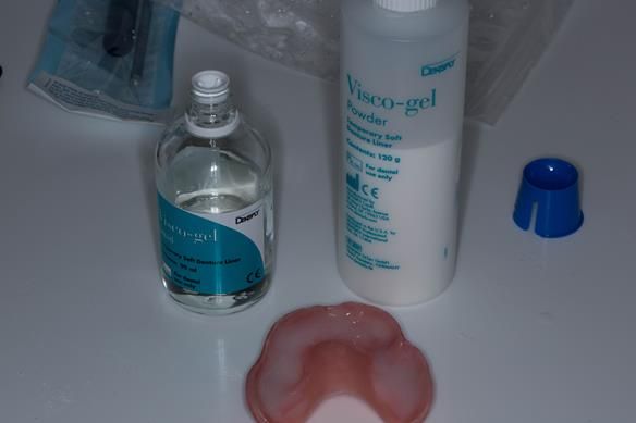
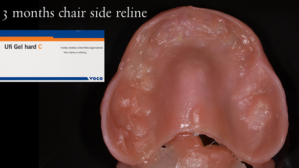
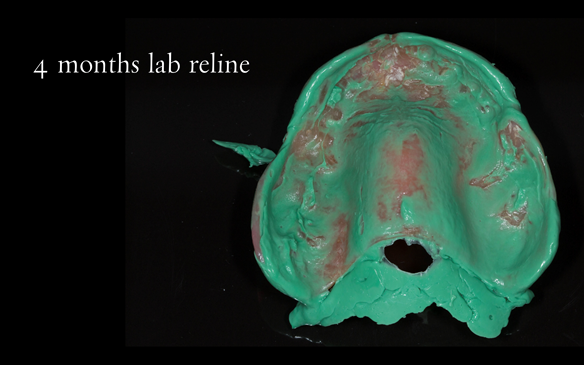
  3. Reviews of the immediate dentures and relines as needed over 9 - 12 months
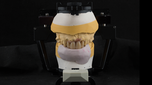
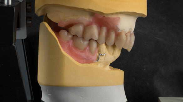
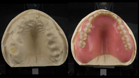
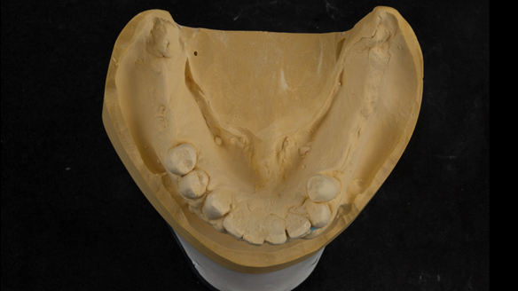
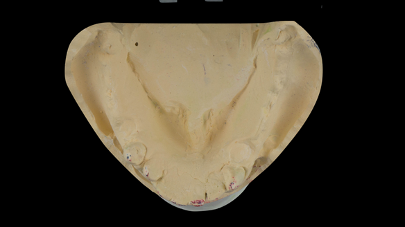
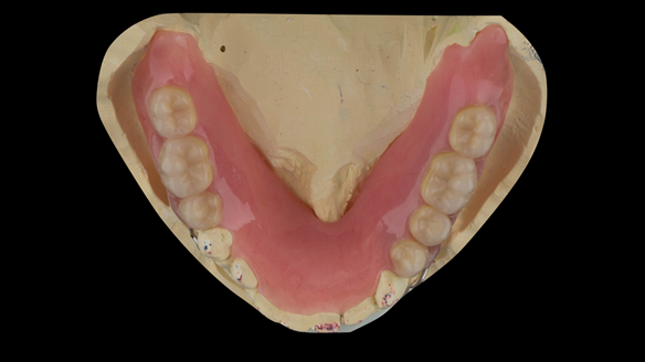
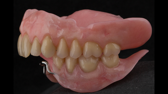
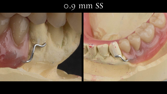
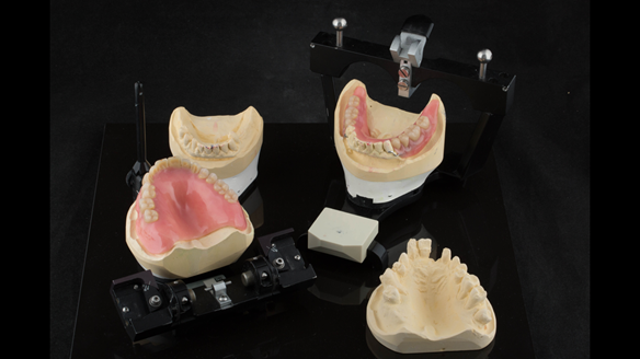
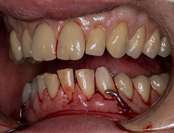
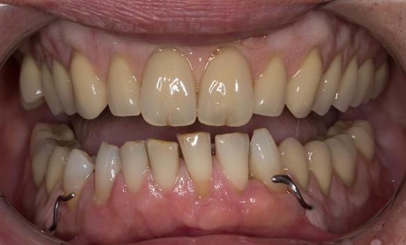
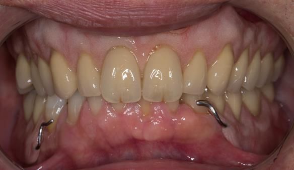
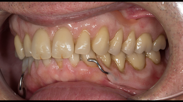
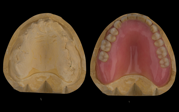
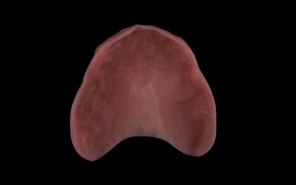
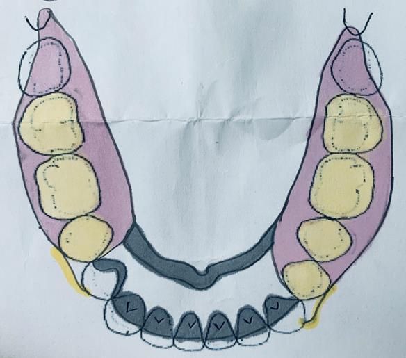
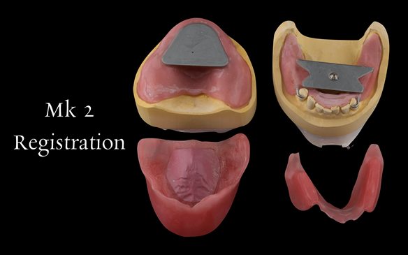
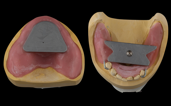
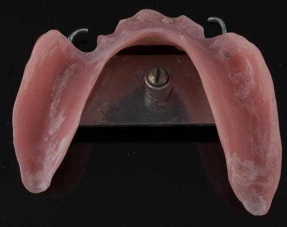
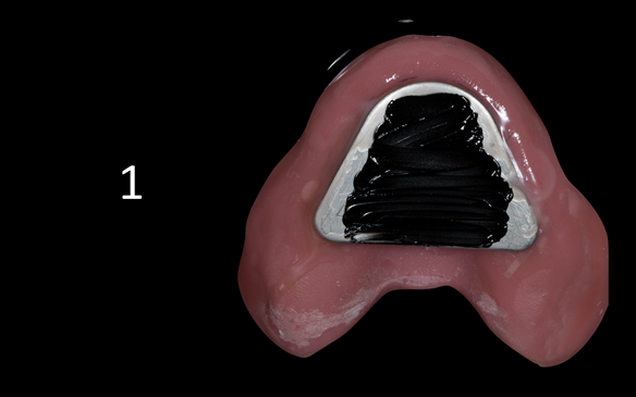
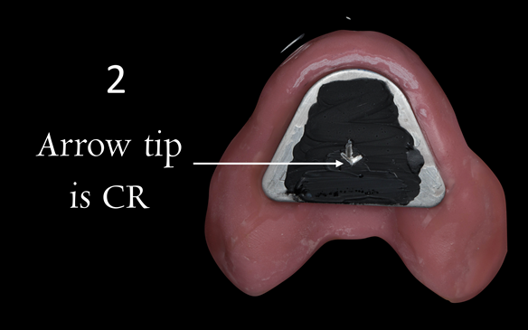
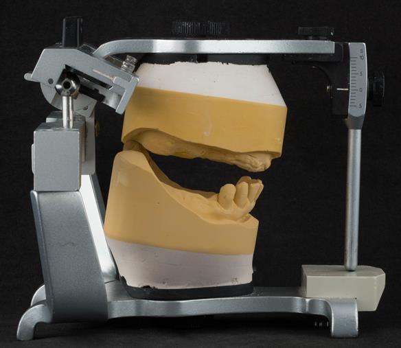
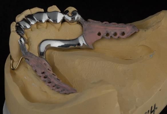
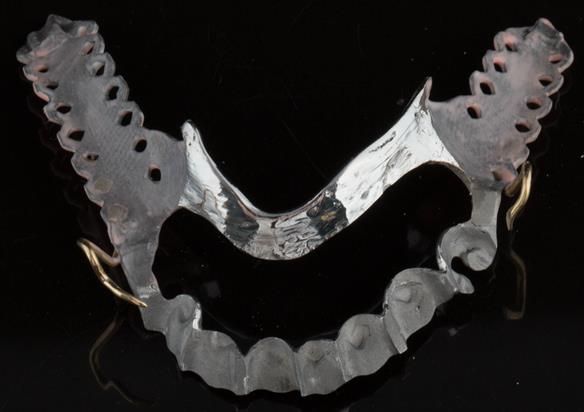
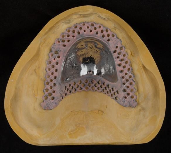
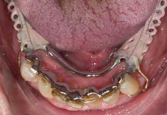
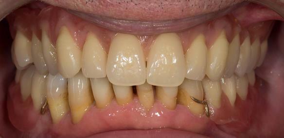
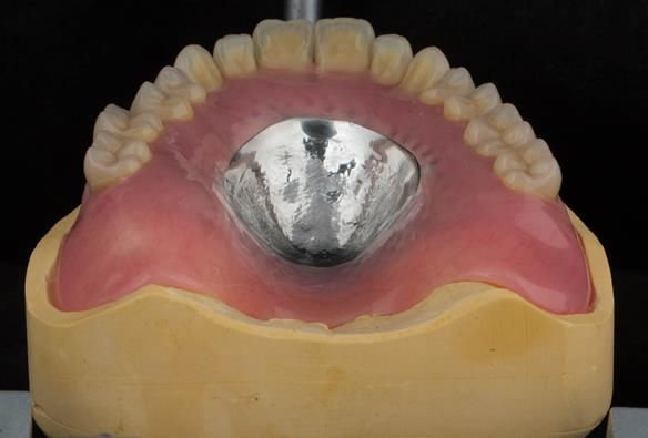
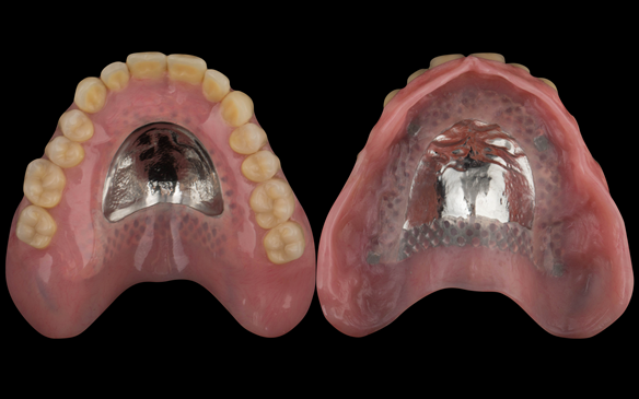
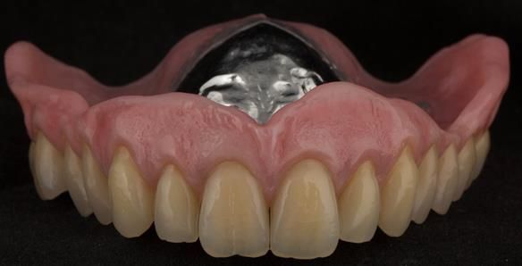
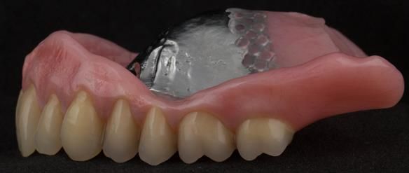
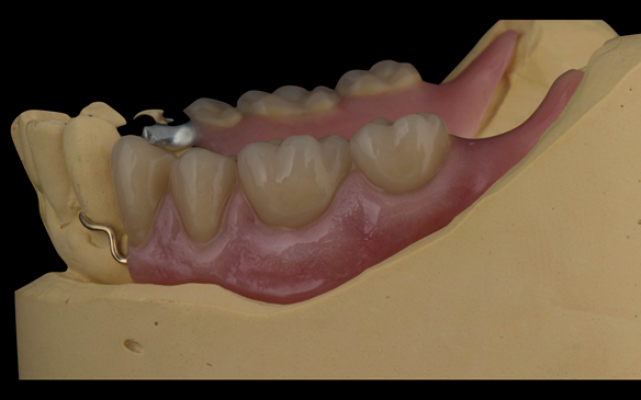
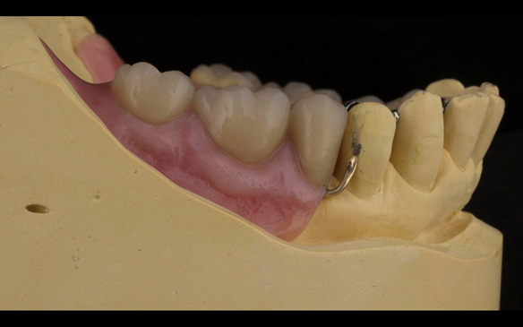
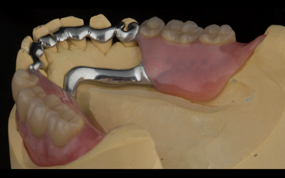
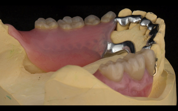
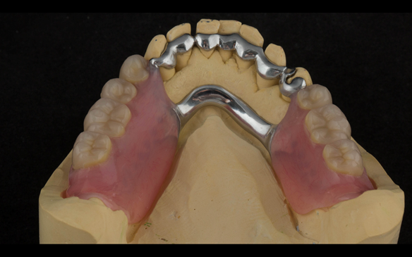
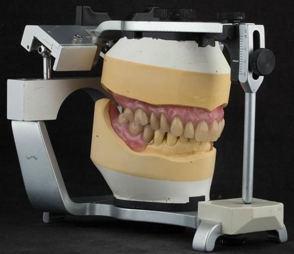
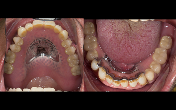
  4. Definitive dentures (Mk 2) – complete upper metal reinforced and lower cobalt chromium based partial of hygienic Scandinavian design to be made 9 - 12 months after extractions of all upper teeth and LR5 and LL4

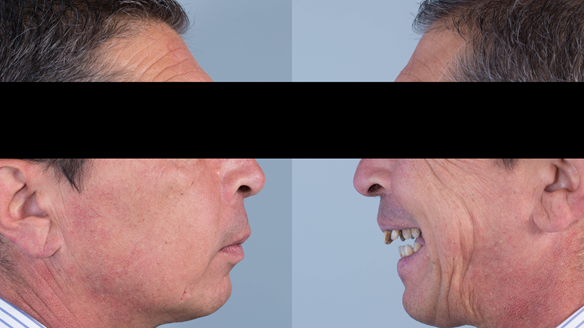
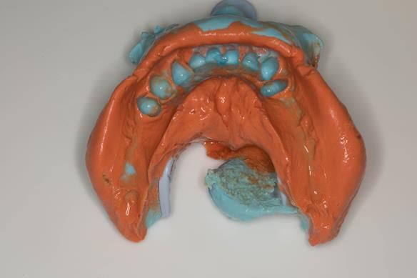
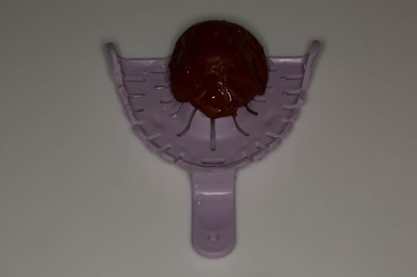
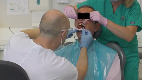
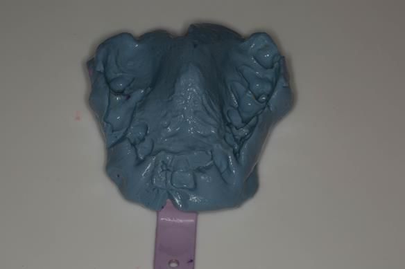
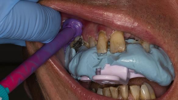
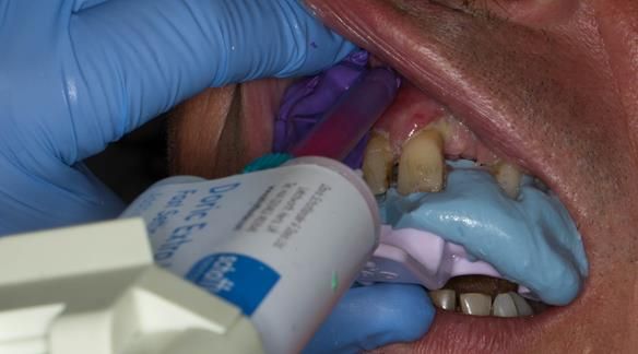
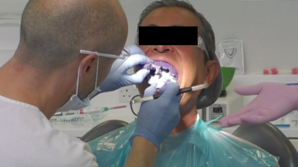
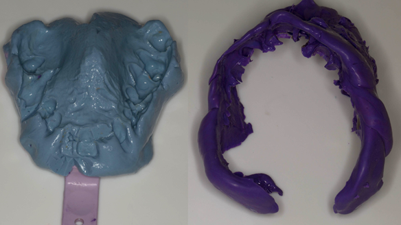
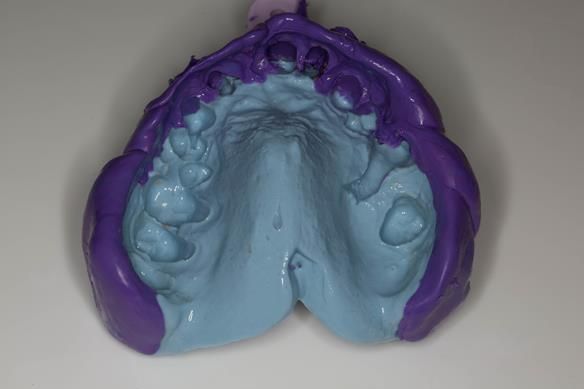
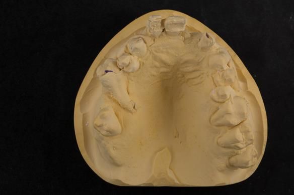
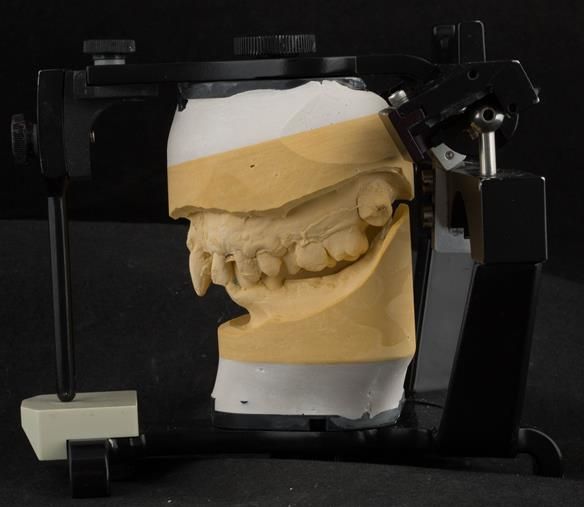
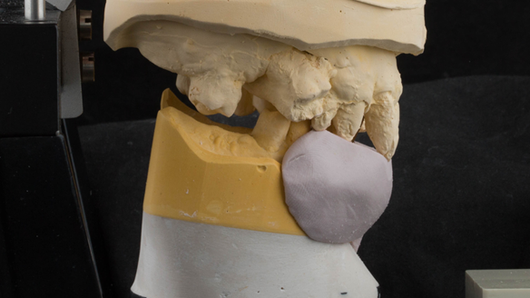
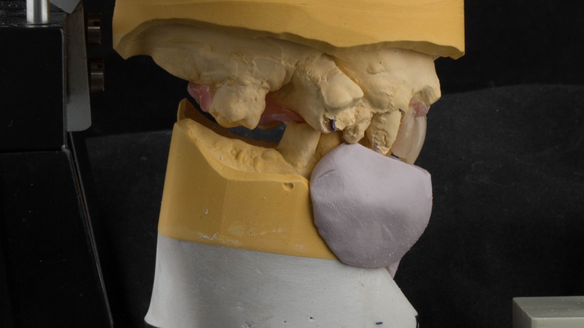
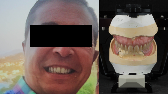
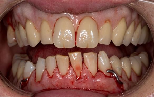
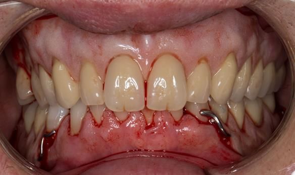
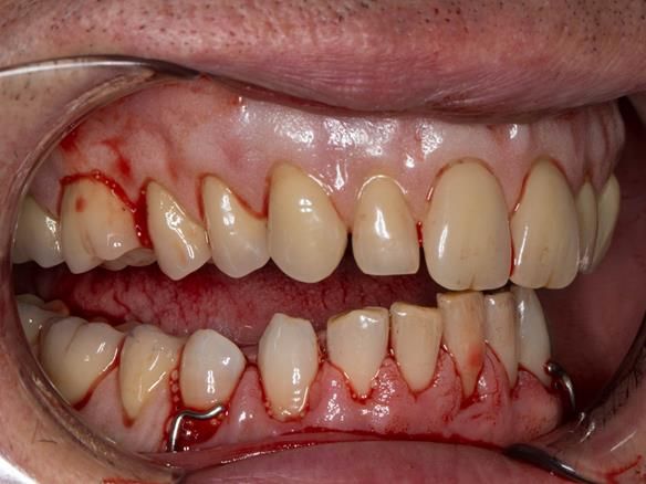
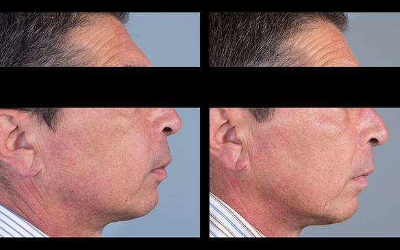
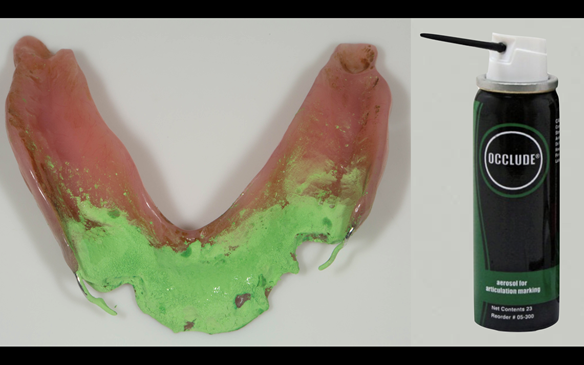
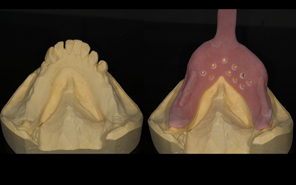
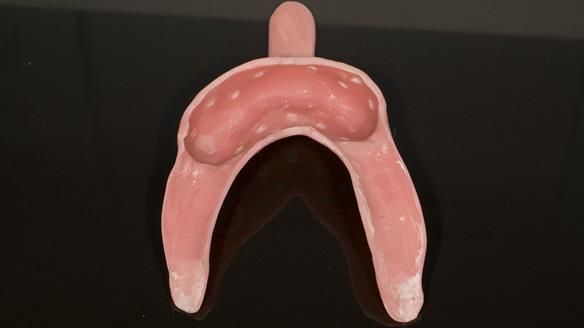
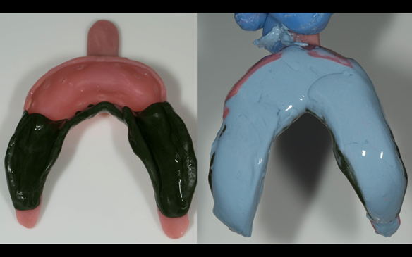
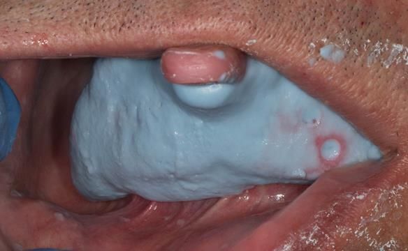
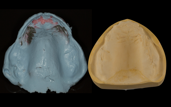
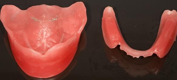
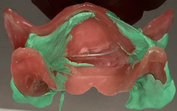
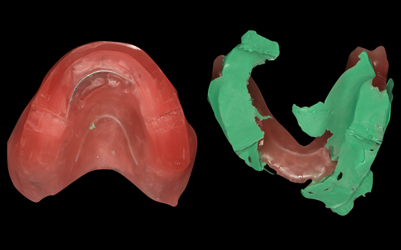
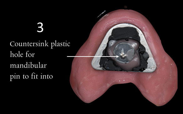
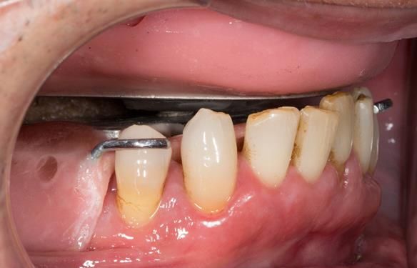
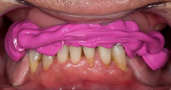
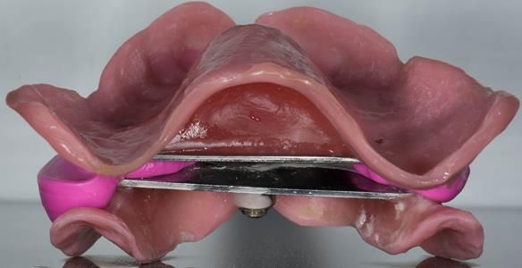
The clinical situation and treatment process is shown in detail below with photographs.

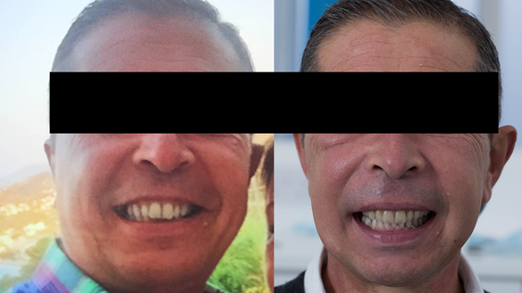
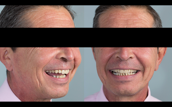
The patient has been successfully rehabilitated and is now having periodontal maintenance from Syed Abad, Specialist in Periodontics at the practice. His quality of life has improved considerably. The clinical prosthodontics was provided by me and the technical work by Rowan.

If you enjoy my Newsletters and you have friends, colleagues, dental students, dental technicians, clinical dental technicians and postgraduate dentists that you think will appreciate them, please feel free to share them. In addition, if your colleagues would like to receive our Newsletters please email me and I will add them to my email list.

Figure 1 Pre-treatment with generalised periodontitis; stage IV grade C, unstable, risk factors: smoker. Prior to phase one treatment of immediate complete upper denture and immediate mandibular acrylic based partial denture.
Figure 2 Pre-treatment with generalised periodontitis; stage IV grade C, unstable, risk factors: smoker. Prior to phase one treatment of immediate complete upper denture and immediate mandibular acrylic based partial denture.
Figure 3 Pre-treatment with generalised periodontitis; stage IV grade C, unstable, risk factors: recent smoker. Prior to phase one treatment of immediate complete upper denture and immediate mandibular acrylic based partial denture.
Figure 4 Pre-treatment with generalised periodontitis; stage IV grade C, unstable, risk factors: smoker. Prior to phase one treatment of immediate complete upper denture and immediate mandibular acrylic based partial denture.
Figure 5 Pre-treatment radiograph indicating bone levels and showing teeth to be removed
Figure 6 treatment planning card containing sequenced treatment plan and quotation. This is how I plan all of my patients treatments
Figure 7 Visit 1. Mandibular primary impression made in two stages using Accudent XD, Ivoclar. This allows full extension to record the sulcus. This was used to produce a primary cast on which the immediate denture was constructed. The teeth were too mobi
Figure 8 Inter-maxillary registration using Futar D bite registration material in intercuspal position (ICP). The Mk 1 immediate complete denture will conform to ICP. Ideally the immediate complete denture would be fabricated in centric relation (CR)
Figure 9 Pre-treatment occlusal view of upper teeth
Figure 10 Modified stock tray with labial wall removed. Red cake compound placed in the palate to aid positioning of the tray
Figure 11 Modified stock tray with labial wall removed. Red cake compound placed in the palate to aid positioning of the tray
Figure 12 Maxillary stock tray impression made in alginate (Dentsply Blueprint) recording the palate, palatal and occlusal surfaces of the teeth. I didn't want to extract the teeth in the impression stage 1
Figure 13 Maxillary stock tray impression made in alginate (Dentsply Blueprint) recording the palate, palatal and occlusal surfaces of the teeth. I didn't want to extract the teeth in the impression - stage 1
Figure 14 Stage 2 primary impression - Alginate reseated in the mouth Schottlander Doric Heavy bodied silicone used to record the depth sulcus
Figure 15 Stage 2 primary impression - Alginate reseated in the mouth Schottlander Doric Heavy bodied silicone used to record the depth sulcus
Figure 16 Careful removal of the impression from the mouth in two pieces - avoiding extraction the mobile teeth
Figure 17 The two parts of the impression ready to fitted back together with superglue (cyanoacrylate adhesive)
Figure 18 The two parts of the impression are fitted back together and glued with superglue (cyanoacrylate adhesive)
Figure 19 Cast for making the immediate complete denture with full depth of the sulcus recorded
Figure 20 Cast for making the immediate complete denture with full depth of the sulcus recorded mounted in intercuspal position for Mk 1 denture. Mk 2 denture was made to centric relation using central bearing appliance
Figure 21 Photos of the patients natural dentition he wanted mimicking
Figure 22 This pre-treatment photo on the right was used as the basis for the Mk 1 immediate denture
Figure 23 Superimposed photograph of the pretreatment smile onto the mounted casts
Figure 24 Mounted casts for Mk 1 denture. Silicone index used as a guide for positioning denture teeth
Figure 25 UR21 UL2 teeth removed from cast and UR21 UL12 teeth arranged to pre-drifted positions
Figure 26 Mounted casts for Mk 1 denture. Silicone index used as a guide for positioning denture teeth
Figure 27 UR21 UL2 teeth removed from cast and UR21 UL12 teeth arranged to pre-drifted positions
Figure 28 The remaining teeth to be extracted removed from casts with artificial teeth arranged to pre-drifted positions
Figure 29 Inflamed soft tissue indicated on the cast
Figure 30 Inflamed soft tissue removed from the cast by me (clinician) as this was swollen soft tissue. Making a flange to fit over this would have bulked the lip out massively. Having it socket fitted instead would result in a lack of peripheral seal and
Figure 31 Finished denture mimicking the natural teeth
Figure 32 Prepared maxillary cast and the finished Mk 1 complete acrylic denture
Figure 33 Unprepared definitive cast for lower Mk 1 denture
Figure 34 Prepared primary mandibular cast. Minimal preparation of the cast apart from careful removal of the teeth to be extracted owing to gross periodontal attachment destruction and alveolar bone loss
Figure 35 Mandibular immediate acrylic based partial denture. Lingual surfaces extended to the mylohyoid line avoiding lingual frenum. Schottlander Enigmalife teeth
Figure 36 Mk 1 immediate acrylic based dentures. Schottlander Enigmalife teeth. 0.9 mm wrought stainless steel clasps on LR4 and LL3
Figure 37 Mandibular immediate acrylic based partial denture with 0.9 mm wrought stainless steel clasps on LR4 and LL3
Figure 38 Mk1 immediate acrylic based dentures ready for fitting at visit 2
Figure 39 Visit two removal of all upper natural teeth, LR5 and LL4
Figure 40 Viscogel reline at fitting (visit 2) appointment
Figure 41 Viscogel reline at fitting (visit 2) appointment
Figure 42 Wax covering the labial aspect of the upper front teeth preventing run of viscogel into the interdental embrasures. This helps speed up clean up of labial surface of the denture. The wax can be pealed off when the viscogel is hard enough.
Figure 43 Visit 2 fitting of Mk 1 upper complete and lower partial immediate dentures
Figure 44 Visit 2 fitting of Mk 1 upper complete and lower partial immediate dentures
Figure 45 Visit 2 fitting of Mk 1 upper complete and lower partial immediate dentures
Figure 46 Visit 2 fitting of Mk 1 upper complete and lower partial immediate dentures
Figure 47 Visit 2 fitting of Mk 1 upper complete and lower partial immediate dentures
Figure 48 Visit 2 fitting of Mk 1 upper complete and lower partial immediate dentures
Figure 49 Visit 3 Review 1 post extraction of Mk 1 upper complete and lower partial immediate dentures
Figure 50 Visit 3 Review 1 post extraction
Figure 51 Visit 3 Review 1 post extraction
Figure 52 Visit 3 Review 1 post extraction
Figure 53 Mid treatment thank you card whilst patient had Mk 1 immediate dentures in situ
Figure 54 3 months post extraction. Mk 1 reline of intaglio surface with Ufi Gel Hard.
Figure 55 Wax covering the labial aspect of the Mk 1 upper front teeth preventing run of Ufi Gel into the interdental embrasures. This helps speed up clean up of labial surface of the denture. The wax can be pealed off when the Ufi Gel is hard enough.
Figure 56 Ufi Gel chair-side reline of upper Mk 1 denture - showing infill of flange UL45 frenum region
Figure 57 4 months post extraction reline impression using light bodied silicone in fitting surface using Doric Flo Light impression material. Firm placement in the mouth to ensure correct seating and border moulding
Figure 58 Dental laboratory reline cast without and with denture
Figure 59 Dental laboratory relined intaglio surface using Pegasus Cold Cure acrylic - Schottlander
Figure 60 5 months post fitting of Mk 1 immediate dentures
Figure 61 5 months post fitting of Mk 1 immediate dentures
Figure 62 12 months post extraction of natural teeth - primary impression lower arch. Part 1 red cake (Kerr) compound to capture saddles and Part 2 impression making with alginate (Dentsply Blueprint)
Figure 63 Maxillary primary impression made in two stages using Accudent XD, Ivoclar. This allows full extension to record the sulcui
Figure 64 Design of metal based lower denture
Figure 65 Sandblasting the lingual of the lower anterior teeth prior to provision of composite rest seats to assist in stabilising and supporting the metal based lower denture
Figure 66 Etching the lingual of the lower anterior teeth prior to provision of composite rest seats to assist in stabilising and supporting the metal based lower denture
Figure 67 Composite rest seats placed on the lingual of the lower anterior teeth to assist in stabilising and supporting the metal based lower denture
Figure 68 Composites after shaping with diamond bur on the lingual of the lower anterior teeth to assist in stabilising and supporting the Mk 2 metal based lower denture
Figure 69 Mk 1 immediate lower partial denture requires adjustment to seat properly over the rest seats using Occlude spray
Figure 70 Mandibular primary cast and special tray for Mk 2 denture
Figure 71 Mandibular special tray (close fitting on saddles spaced over the teeth with perforations) for Mk 2 denture
Figure 72 greenstick on saddles border moulded. Adhesive placed (blueprint) alginate mixed, glazed with wet finger and ready to take the impression
Figure 73 Definitive impression in situ after border moulding
Figure 74 Mandibular definitive impression made in alginate
Figure 75 Maxillary spaced special tray for alginate
Figure 76 Maxillary special tray border moulded with greenstick compound, ensuring a peripheral seal is obtained.
Figure 77 Maxillary special tray prepared with alginate adhesive
Figure 78 Definitive impression made in alginate (Blueprint) with resultant poured definitive cast
Figure 79 Primary registration rims used to record the APPROXIMATE centric relation (CR) and occlusal vertical dimension (OVD) to enable mounting of the definitive casts to fabricate the central bearing apparatus plates parallel to each other. Important s
Figure 80 Primary registration rims used to record the APPROXIMATE centric relation (CR) and occlusal vertical dimension (OVD) to enable mounting of the definitive casts to fabricate the central bearing apparatus plates parallel to each other. Important s
Figure 81 Primary registration rims used to record the APPROXIMATE centric relation (CR) and occlusal vertical dimension (OVD) to enable mounting of the definitive casts to fabricate the central bearing apparatus plates parallel to each other. Important s
Figure 82 Visit 3 for Mk 2 fabrication. Registration visit with central bearing apparatus for CR recording and wax rims for tooth position and OVD recording
Figure 83 Visit 3 for Mk 2 fabrication. Inter-maxillary registration with central bearing apparatus made on light cured tray maker for accurate CR recording
Figure 84 Visit 3 for Mk 2 fabrication. Inter-maxillary registration with central bearing apparatus made on light cured tray maker for accurate CR recording - mandibular fitting surface
Figure 85 Visit 3 for Mk 2 fabrication. Inter-maxillary record of the maxillary Mk 1 denture. This allowed a copy of the Mk 1 to be mounted on the articulator to allow the Mk 2 denture to mimic the tooth arrangement. The patient was happy with the aesthet
Figure 86 Visit 3 for Mk 2 fabrication. Inter-maxillary record of the maxillary Mk 1 denture. This allowed a copy of the Mk 1 to be mounted on the articulator to allow the Mk 2 denture to mimic the tooth arrangement. The patient was happy with the aesthet
Figure 87 Visit 3 for Mk 2 fabrication. Inter-maxillary record of the maxillary Mk 1 denture. This allowed a copy of the Mk 1 to be mounted on the articulator to allow the Mk 2 denture to mimic the tooth arrangement. The patient was happy with the aesthet
Figure 88 Visit 3 for Mk 2 fabrication. Inter-maxillary registration with central bearing apparatus made on light cured tray maker for accurate CR recording - at the OVD of Mk 1
Figure 89 Visit 3 for Mk 2 fabrication. Inter-maxillary registration with central bearing apparatus. Maxillary plate with china graph pencil marking for scribing the CR arrow head
Figure 90 Visit 3 for Mk 2 fabrication. Inter-maxillary registration with central bearing apparatus. Maxillary plate with china graph pencil marking with arrow head scribed showing precise CR
Figure 91 Visit 3 for Mk 2 fabrication. Inter-maxillary registration with central bearing apparatus. Maxillary plate with plastic disc with countersunk hole placed over CR arrow head. This allows the mandibular pin to fit into the hole to enable accurate
Figure 92 Visit 3 for Mk 2 fabrication. Inter-maxillary registration with central bearing apparatus in the mouth with the maxillary and mandibular parts "locked" together in CR by the pin in the hole
Figure 93 Visit 3 for Mk 2 fabrication. Inter-maxillary registration with central bearing apparatus in the mouth with the maxillary and mandibular parts registered together with futar D bite registration material
Figure 94 Visit 3 for Mk 2 fabrication. Inter-maxillary registration with central bearing apparatus with the maxillary and mandibular parts registered together with futar D bite registration material enabling mounting of maxillary and mandibular definitiv
Figure 95 Mounted maxillary and mandibular definitive casts at desired OVD using the wax rim with facebow and in CR via the central bearing apparatus
Figure 96 Mandibular cobalt chromium based framework (Chris Hesketh) - opaqued saddles - Scandinavian hygienic design with 0.9 mm wrought gold clasps on LR4 and LL3
Figure 97 Mandibular cobalt chromium based framework (Chris Hesketh) - opaqued saddles - Scandinavian hygienic design showing composite rest seat indentations on the lingual surfaces of the frame with 0.9 mm wrought gold clasps on LR4 and LL3
Figure 98 Cobalt chromium base for complete upper denture for increased fracture resistance (Chris Hesketh). This will have an acrylic post dam which I find results in increased suction (retention) and enables relining in the future should this be needed
Figure 99 Cobalt chromium framework trial insertion in the mouth. The lingual bar does not rest on the soft tissues - blocked by a wash of wax less than 0.5mm thick
Figure 100 Mk 2 mandibular denture try in - teeth in wax
Figure 101 Mk 2 dentures try in - teeth in wax assessment of tooth positions and occlusion
Figure 102 Mk 2 definitive denture try in - at this visit the patient assesses the aesthetics by 1. video, 2. still photographs comparing Mk 1 with Mk 2 and direct observation in a mirror. Only when he was completely happy do we proceed with finishing.
Figure 103 Mk 2 maxillary based denture with acrylic post dam which I find results in increased suction (retention) and enables relining in the future should this be needed
Figure 104 Mk 2 maxillary based denture with acrylic post dam which I find results in increased suction (retention) and enables relining in the future should this be needed
Figure 105 Mk 2 maxillary metal based denture with subtlety characterised Schottlander Enigmalife teeth. Gum colouring - Vertex
Figure 106 Mk 2 maxillary metal based denture with subtlety characterised Schottlander Enigmalife teeth. Gum colouring - Vertex
Figure 107 Mk 2 maxillary metal based denture with subtlety characterised Schottlander Enigmalife teeth. Gum colouring - Vertex
Figure 108 Mk 2 mandibular metal based denture with Schottlander Enigmalife teeth. Gum colouring - Vertex with 0.9 mm wrought gold clasps on LR4 and LL3
Figure 109 Mk 2 mandibular metal based denture with Schottlander Enigmalife teeth. Gum colouring - Vertex with 0.9 mm wrought gold clasps on LR4 and LL3
Figure 110 Mk 2 mandibular metal based denture showing open Scandinavian hygienic design.
Figure 111 Mk 2 mandibular metal based denture showing open Scandinavian hygienic design.
Figure 112 Mk 2 mandibular metal based denture showing open Scandinavian hygienic design.
Figure 113 mandibular metal based denture showing fitting surface. Polished bar over the soft tissues. Not polished on tooth support increasing retention
Figure 114 Mk 2 finished dentures on the articulator
Figure 115 12 months after removal of failing teeth. During this time Mk 1 immediate dentures were fitted, periodontal therapy (including periodontal flap surgery) was provided
Figure 116 Mk 2 finished dentures fitted with 0.9 mm wrought gold clasps on LR4 and LL3
Figure 117 Mk 2 finished dentures fitted with 0.9 mm wrought gold clasps on LR4 and LL3
Figure 118 Mk 2 finished dentures fitted
Figure 119 Before treatment and after with Mk 2 maxillary cobalt chromium based complete denture and mandibular cobalt chromium based partial denture fitted
Figure 120 Before treatment and after treatment with Mk 2 maxillary cobalt chromium based complete denture and mandibular cobalt chromium based partial denture fitted. Periodontal therapy including surgery by Syed Abad, Specialist in Periodontics.

In-House courses for 2020

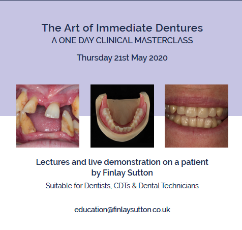
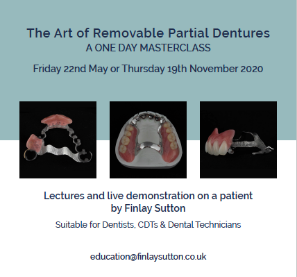
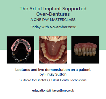
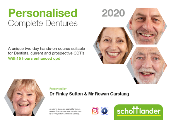
Finlay's in-house denture courses with PDF application forms:

Please click on the images below.

Full Protocol - Transition through extractions and acrylic based immediate dentures to metal based definitive dentures

Reference material

Full access PDF to my published scientific papers which explain my philosophy and clinical techniques. Please click on the link below and scoll down this page to find lots of useful clinical techniques, reference material and previous lectures:

https://www.finlaysutton.co.uk/speaking

Massive suction on lower complete denture - full treatment walk through by Finlay Sutton
One to One Removable Prosthodontics Denture Mentorship with Finlay
Finlay designs a Kennedy class 1 upper metal based partial denture for Sue in real time.
Finlay designs a Kennedy class 2 (1) metal based partial denture for Sylvia - who has had MRONJ - VIDEO ON YouTube

Finlay's Blog

Massive suction on lower complete denture - full treatment walk through by Finlay Sutton

Welcome to my Newsletter 62 video walk through, where I show the making and fitting of complete dentures plus extractions for Rafique. This issue provides a comprehensive overview of the entire protocol workflow.

One to One Removable Prosthodontics Denture Mentorship with me, Finlay

Hi there! I'm here to help you achieve the best results with removable pros. I can assist with complete dentures, interesting partials, and even implants over dentures. Let's work together to reduce reviews, make patients happier, and provide better outcomes. If you need mentorship, coaching, or treatment planning, reach out to me at education@finlaysutton.com.

The cost for mentorship is £450 per hour, plus VAT.

Finlay designs a Kennedy class 1 upper metal based partial denture for Sue in real time

Finlay designs a Kennedy class 1 upper metal based partial denture for Sue in real time.

This is how I design RPDs for all of my cases. YouTube Video.

Finlay designs a Kennedy class 2 (1) metal based partial denture for Sylvia - who has had MRONJ - VIDEO ON YouTube

Finlay designs a Kennedy class 2 (1) metal based partial denture for Sylvia - who has had MRONJ - VIDEO ON YouTube

prev
next