Provision of a maxillary cobalt chromium based partial denture/protective occlusal splint in a heavily restored dentition

Diagnoses:

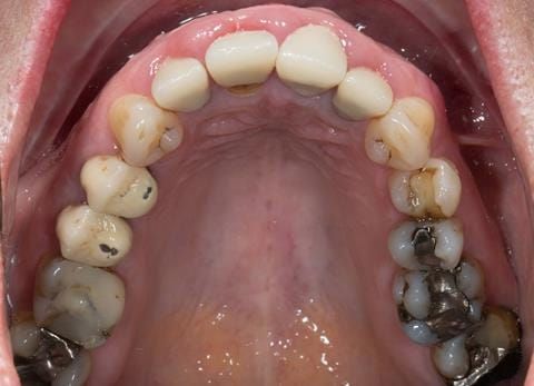
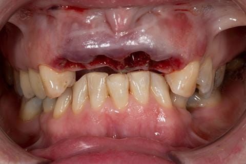
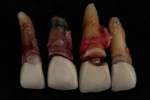
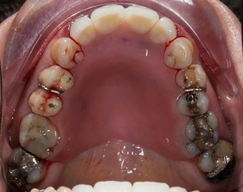
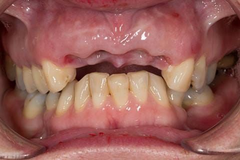
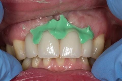
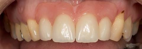
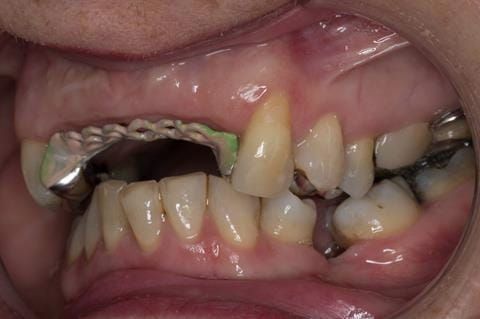
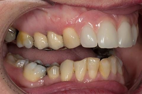
  1. UR2 peri-radicular periodontitis with a peri-radicular area on the root apex. Retrograde amalgam filling from a previous apicectomy. No visible root canal or root canal filling. Large circumferential marginal gap between the crown and tooth. Large post and core present. Very little tooth structure remaining resulting in a fragile tooth with increased potential for fracture.
  2. UR1 peri-radicular periodontitis with a small peri-radicular area on the root apex with wide blunderbuss apex. Radio-opaque root canal filling present approximately 3 mm short of the radiographic apex. Large circumferential marginal gap between the crown and tooth. Large post and core present. Very little tooth structure remaining resulting in a fragile tooth with increased potential for fracture.
  3. UL1 peri-radicular periodontitis with a peri-radicular area on the root apex. Retrograde amalgam filling from a previous apicectomy. Visible root canal space with no sign of root canal filling. Large circumferential marginal gap between the crown and tooth. Large post and core present. Very little tooth structure remaining resulting in a fragile tooth with increased potential for fracture.
  4. UL2 peri-radicular periodontitis with a peri-radicular area on the root apex. No visible root canal or root canal filling. Large circumferential marginal gap between the crown and tooth. Large post and core present. Very little tooth structure remaining resulting in a fragile tooth with increased potential for fracture.
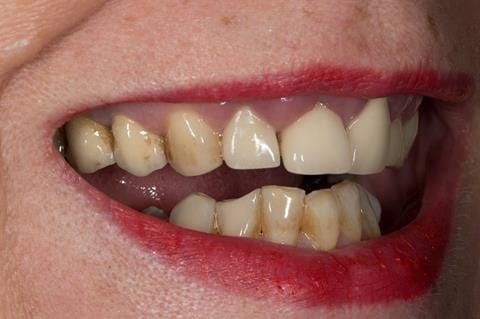
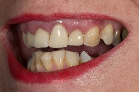
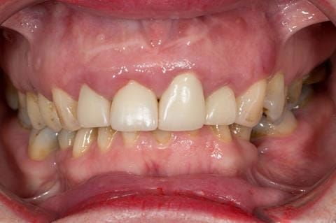
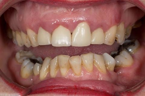
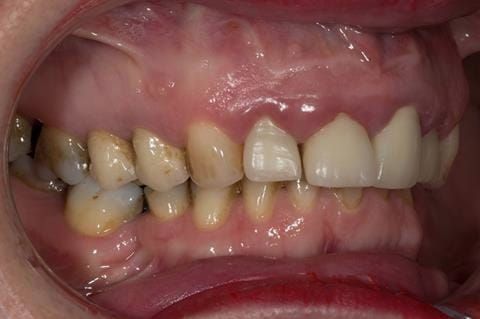
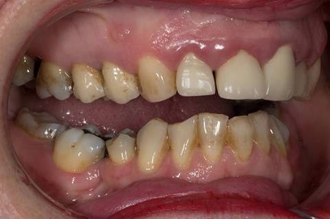
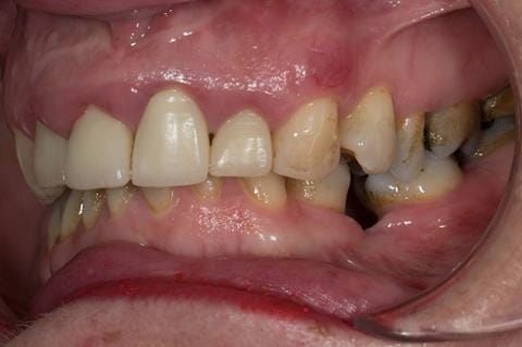
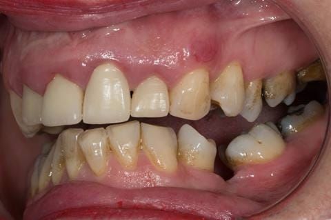
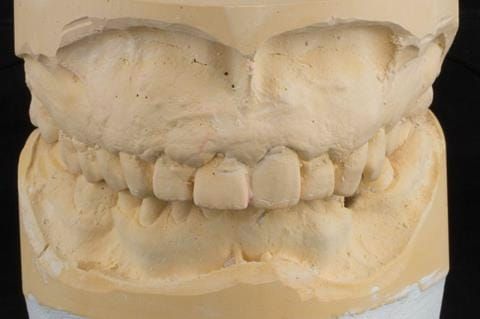
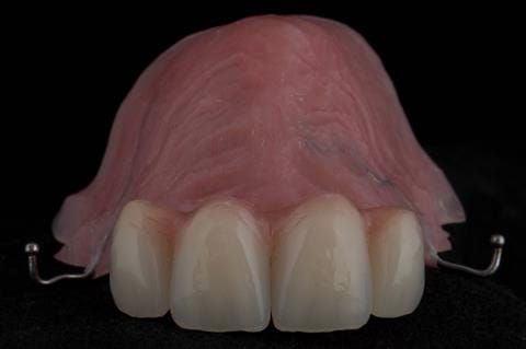
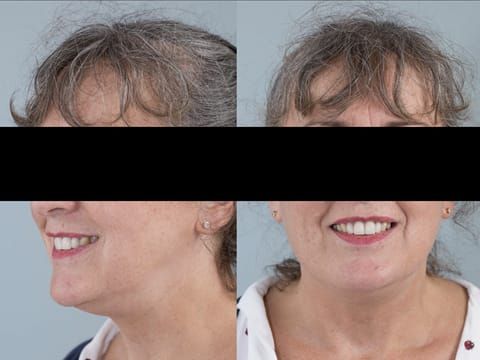
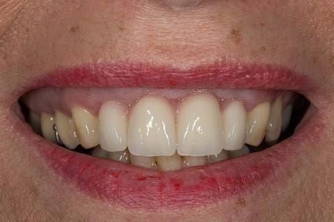
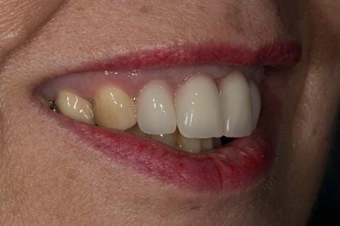
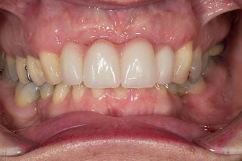
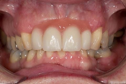
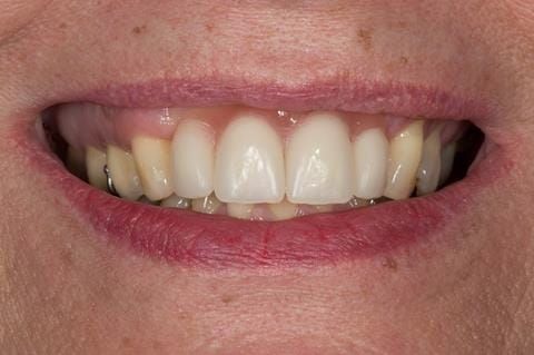
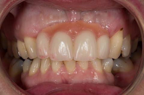
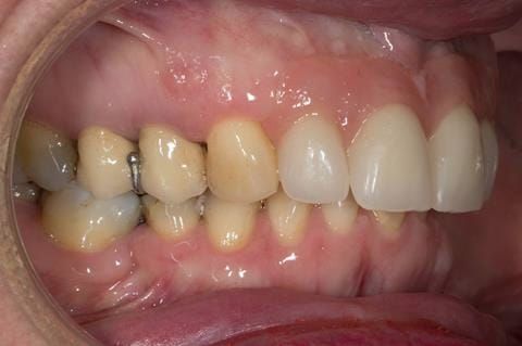
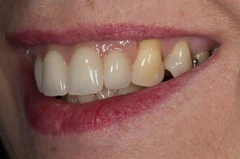
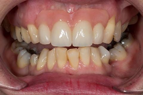
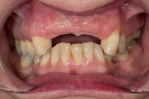
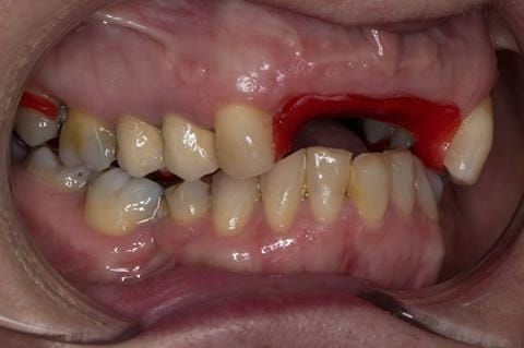
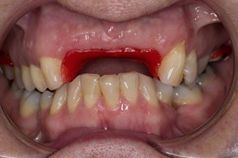
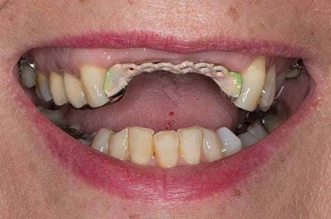
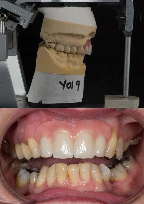
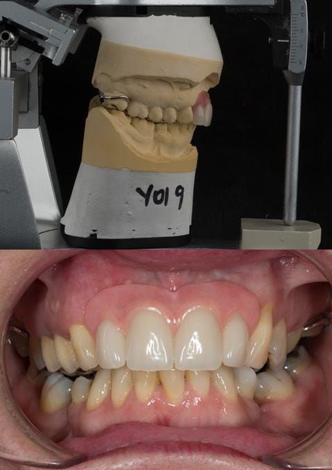
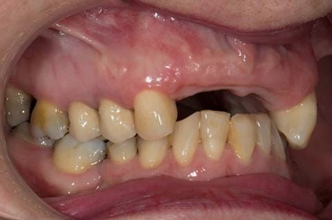
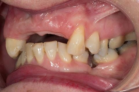
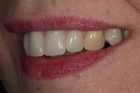
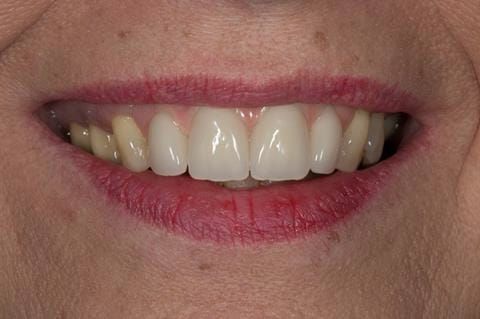
  5. High smile line showing gum above gingival zeniths of upper front teeth when smiling. Aesthetic failure of the upper four incisors with inflammation of the gingivae and mis-match of the gingival zenith levels.
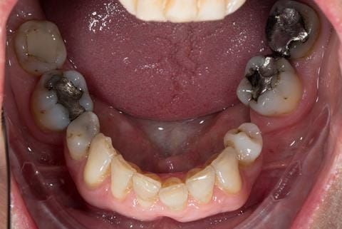
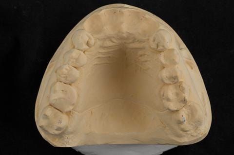
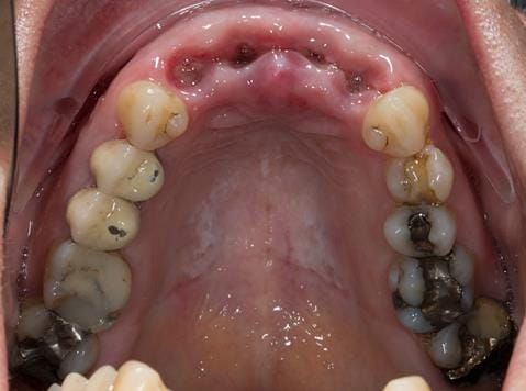
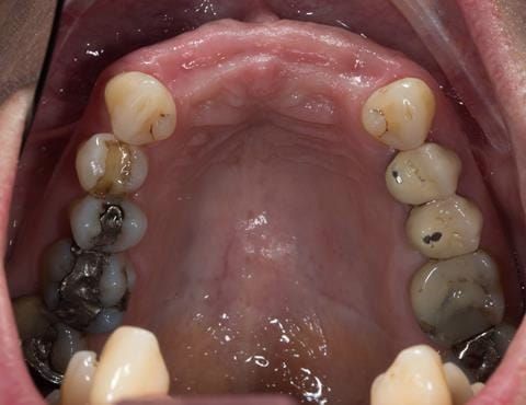
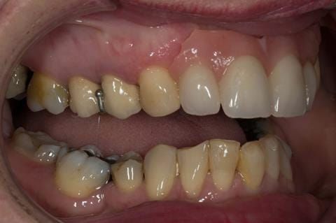
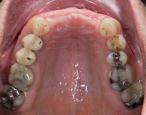
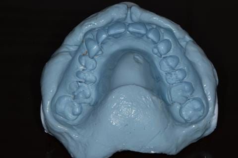
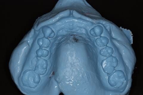
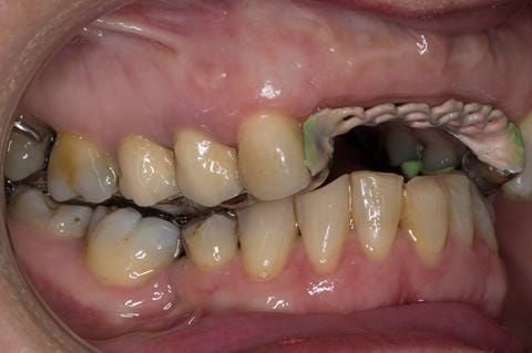
  6. Other than the maxillary incisors the remaining dentition was in marginally better condition being moderately to heavily restored. Many will probably require replacement and restoration from time to time mainly from wear and tear owing to occlusal forces.

Treatment options discussed with the patient:

  1. Do nothing. The patient did not want this option.
  2. Strip down the upper anterior teeth to investigate their restorability, root canal treat as necessary, place new post and cores, fit well adapted provisional crowns, muco-gingival surgery to level the gingival margin zeniths and place definitive crowns. One or more of these teeth may not be restorable - if so replace with a dental implant supported crown (s). The patient did not want to have this option as she did not want to risk future failure of the restored teeth.
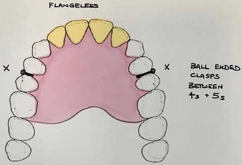
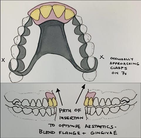
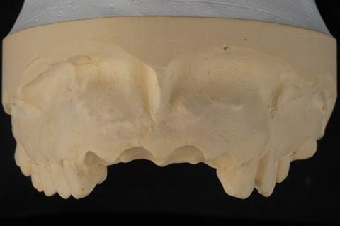
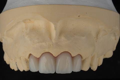
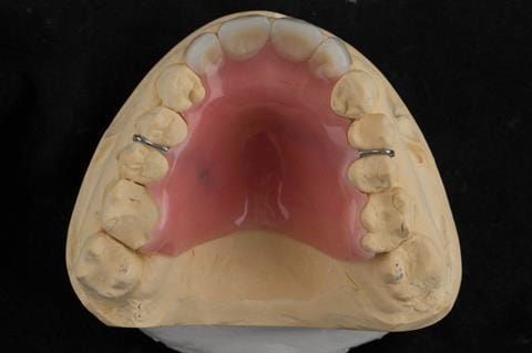
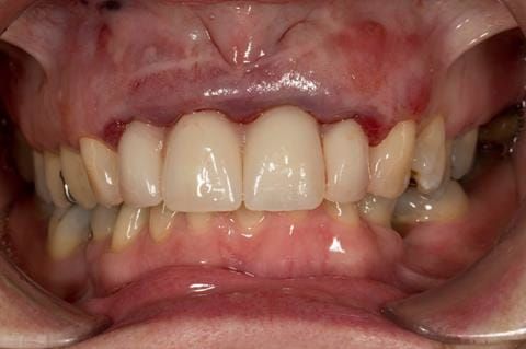
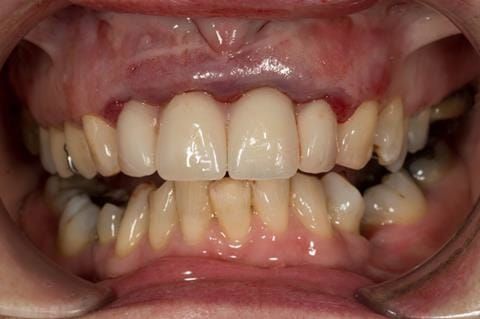
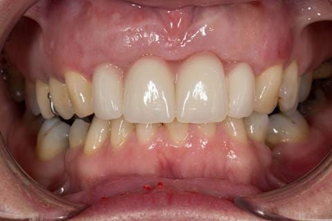
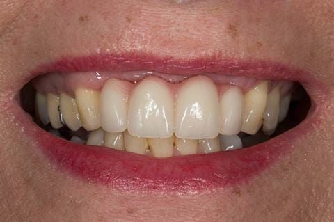
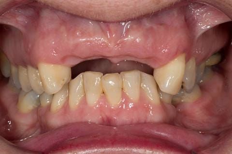
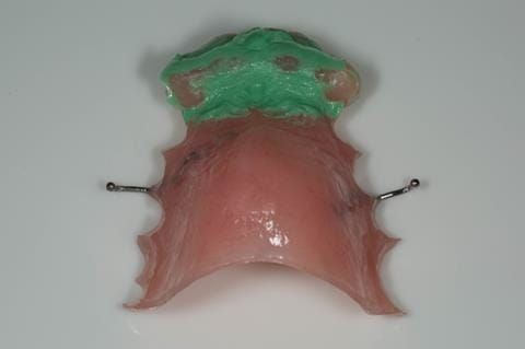
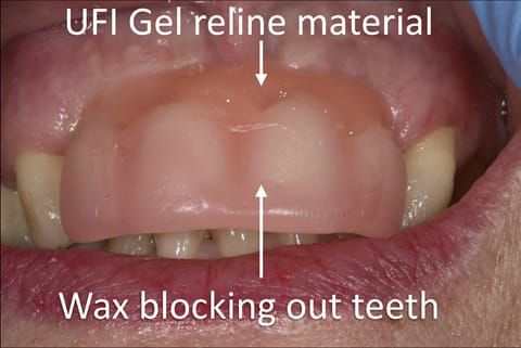
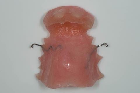
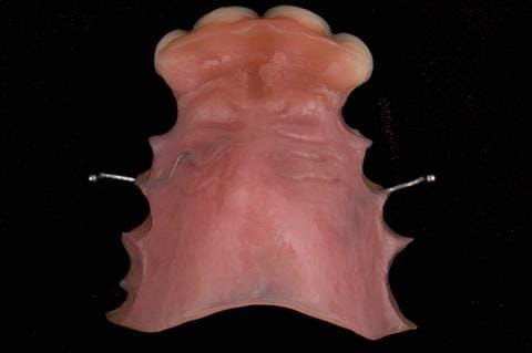
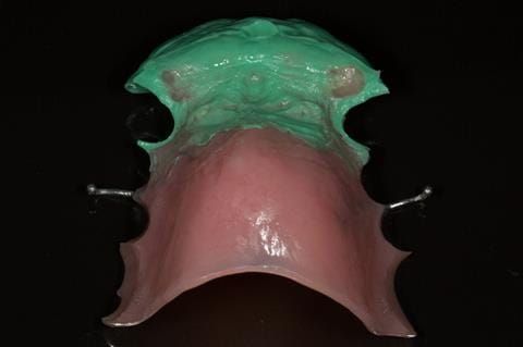
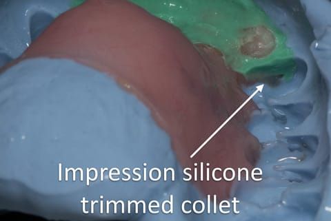
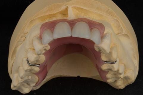
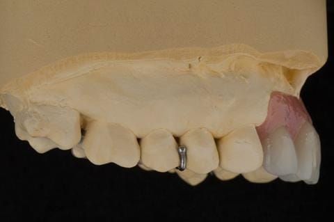
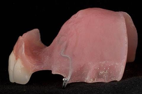
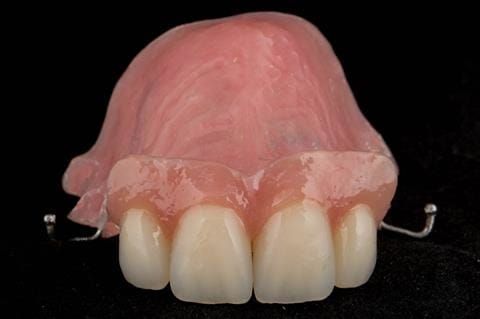
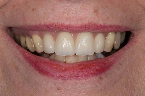
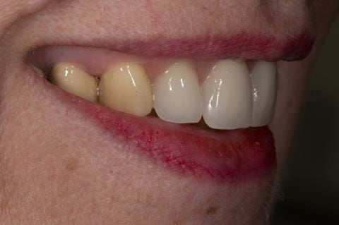
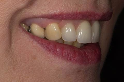
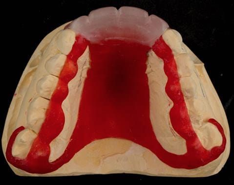
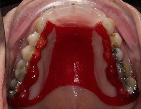
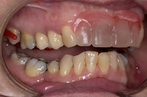
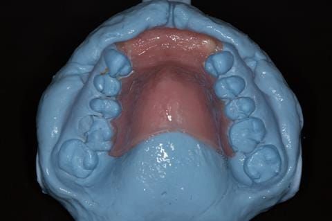
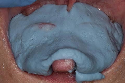
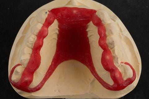
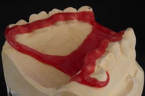
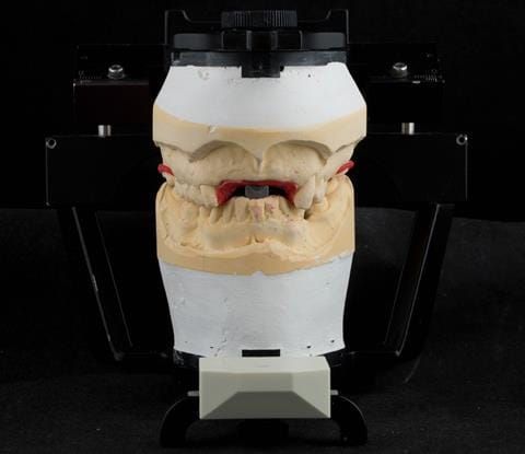
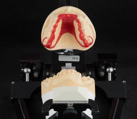
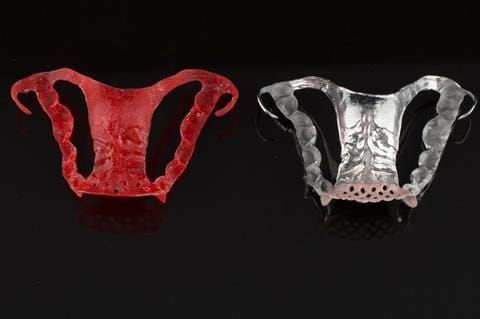
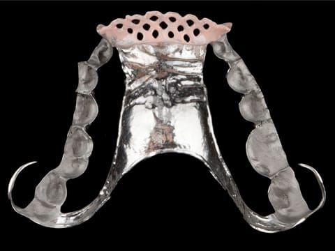
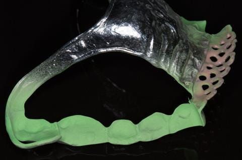
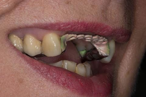
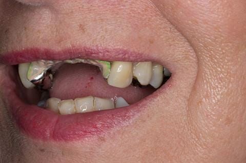
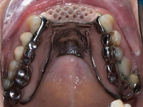
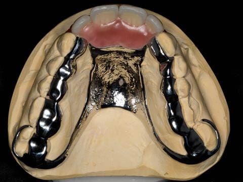
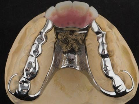
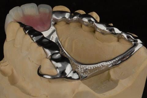
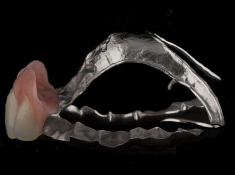
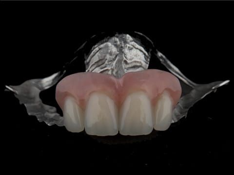
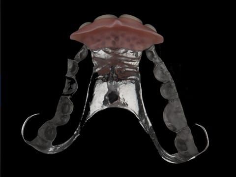
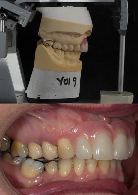
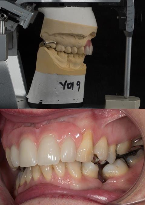
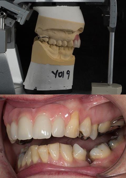
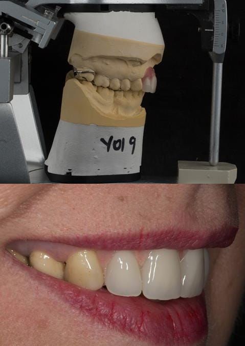
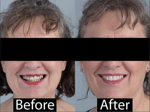
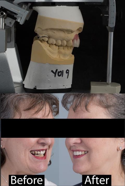
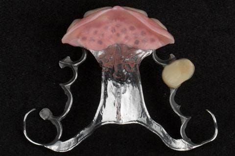
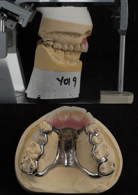
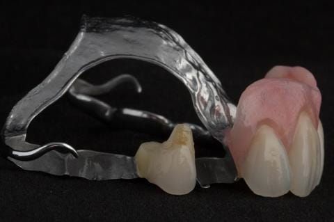
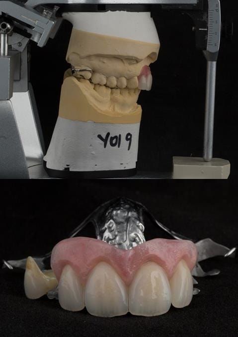
  3. Extract the upper 2-2 teeth and replace with an interim acrylic based partial denture. Reline the interim denture over 9 - 12 months, replacing with a  definitive cobalt chromium based partial denture. The definitive denture would ideally be designed as an occlusal protective splint to reduce the the potential for mechanical wear and breakages of the moderately/heavily restored maxillary dentition. In addition, should further upper teeth require extraction they could be added on to the denture cobalt chromium framework - therefore a new prosthesis would not be required as future teeth are lost. This option would produce an excellent aesthetic outcome. This is the option the patient chose to have.
  4. Extract the upper 2-2 teeth and replace with dental implant supported restorations in the form of bridge or individual crowns. The patient did not wish to have this option owing to financial constraints.

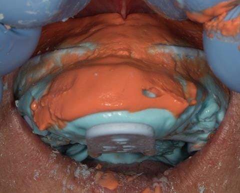
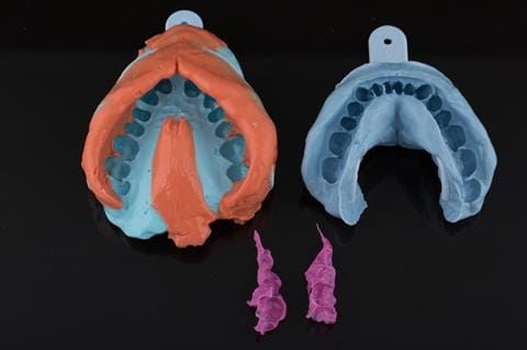
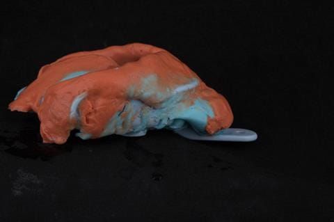
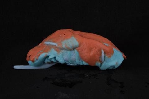
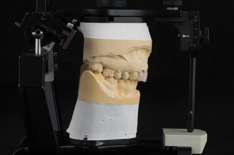
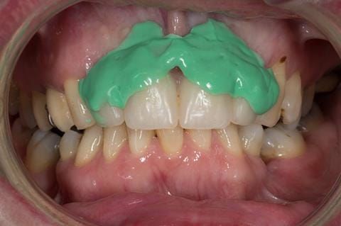
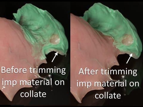
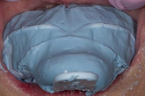
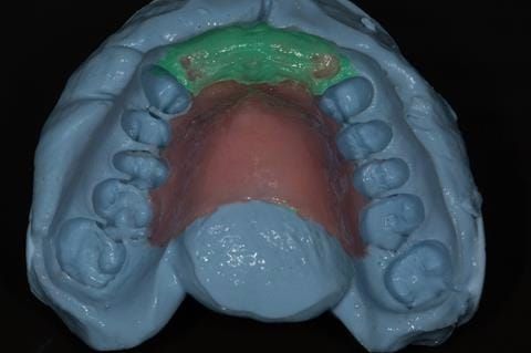
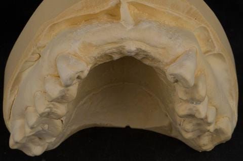
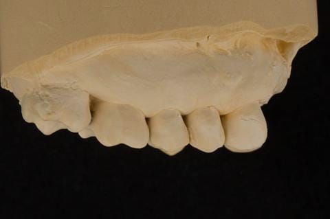
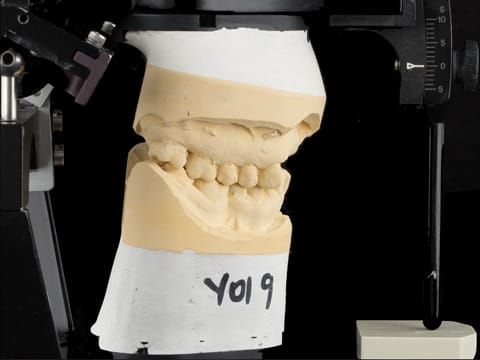
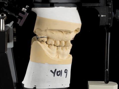
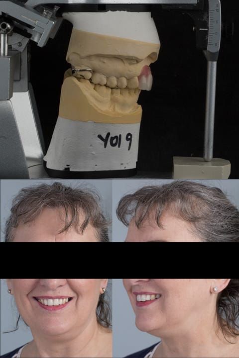
Following consultation and second discussion appointment the patient chose to have option 3 namely, a maxillary cobalt chromium based partial denture/protective occlusal splint. The clinical situation and treatment process is shown in detail below with photographs. The patient was successfully rehabilitated with this and her quality of life considerably improved. The clinical work was provided by Finlay and the technical work by Rowan.

Figure 1 a & b. Pre-treatment showing high smile line and aesthetically poor upper incisors
Figure 2. Pre-treatment showing high smile line and aesthetically poor upper incisors
Figure 3. Pre-treatment showing high smile line and aesthetically poor upper incisors
Figure 4. Pre-treatment showing high smile line and aesthetically poor upper incisors
Figure 5. Pre-treatment - upper four incisors with inflammation of the gingivae and mis-match of the gingival levels.
Figure 6. Pre-treatment - upper four incisors with inflammation of the gingivae and mis-match of the gingival levels.
Figure 7. Pre-treatment - upper four incisors with inflammation of the gingivae and mis-match of the gingival levels.
Figure 8. Pre-treatment - upper four incisors with inflammation of the gingivae and mis-match of the gingival levels.
Figure 9. Pre-treatment - upper four incisors with inflammation of the gingivae and mis-match of the gingival levels.
Figure 10. Pre-treatment - upper four incisors with inflammation of the gingivae and mis-match of the gingival levels.
Figure 11. Pre-treatment - occlusal view - upper arch
Figure 12. Pre-treatment - occlusal view - lower arch.
Fig 13. Pre-treatment radiographs. Maxillary incisors with peri-radicular areas. UR2 UL1 retrograde amalgam fillings from previous apicectomies. Large circumferential marginal gaps between the crowns and teeth on UR12, UL12, large posts and cores present.
Figure 14. Immediate maxillary denture flangeless design, to be fitted at the extraction appointment of UR21, UL12. Colletted with two ball ended clasps. Denture to be worn over 9 - 12 months duration to allow for ridge resorption.
Figure 15. Design of definitive maxillary cobalt chromium based partial denture. Support derived from the remaining natural teeth. The heavily restored posterior dentition to be protected by partial occlusal coverage in metal.
Figure 16. Maxillary primary impression made in two stages using Accudent XD, Ivoclar. This allows full extension to record the sulcus.
Figure 17. Maxillary primary impression made in two stages using Accudent XD, Ivoclar. The mandibular impression made using Blueprint, Dentsply. Inter-maxillary record made in Futar D, Kettenbach.
Figure 18. Maxillary primary impression made in two stages using Accudent XD, Ivoclar. This overextends the impression and captures the full depth of the sulcus. Please note this is from a different patient as I did not photograph the primary impressions.
Figure 19. Maxillary primary impression made in two stages using Accudent XD, Ivoclar. This overextends the impression and captures the full depth of the sulcus. Please note this is from a different patient as I did not photograph the primary impressions.
Figure 20. Duplicate of the primary cast of the maxillary arch showing good extension - enabling the construction of acrylic based immediate partial denture.
Figure 21. Primary cast of the maxillary arch with upper incisors removed and prepared for ovate pontics.
Figure 22. Maxillary cast with immediate partial denture processed and fitted. Schottlander Enigmalife teeth.
Figure 23. Primary cast of the maxillary arch with upper incisors removed and prepared for ovate pontics and post dam scored into the model.
Figure 24. Maxillary cast with immediate partial denture processed and fitted as per design in figure 13. Schottlander Enigmalife teeth.
Figure 25. Maxillary immediate partial denture finished with ovate pontics and ball ended clasps on Denar Mk 2 articulator.
Figure 26. Maxillary immediate partial denture finished. When fitting I always adjust below area where the denture touches the tooth as in the photograph. This prevents a gap forming between the denture and the teeth when fitted - as shown in Figure 32.
Figure 27. Maxillary immediate partial denture finished with ovate pontics and ball ended clasps. Schottlander Enigmalife teeth.
Figure 28. Extraction sites of upper four incisors
Figure 29. Extracted upper four incisors
Figure 30. Maxillary immediate denture fitted just after extraction in shown intercuspal position. Schottlander Enigmalife teeth.
Figure 31. Maxillary immediate denture fitted just after extractionwith teeth apart. Schottlander Enigmalife teeth.
Figure 32. Maxillary immediate denture fitted at the extraction appointment - showing no gaps between the denture and teeth.
Figure 33. Maxillary immediate denture fitted one week after the extraction appointment. Schottlander Enigmalife teeth.
Figure 34. Maxillary immediate denture fitted one week after the extraction appointment. Schottlander Enigmalife teeth.
Figure 35. Maxillary immediate denture fitted one week after the extraction appointment.
Figure 36. Healing sockets at 1 week review.
Figure 37. Maxillary immediate denture fitted one week after the extraction appointment. Schottlander Enigmalife teeth.
Figure 38. Healing sockets at 1 week review.
Figure 39. Maxillary immediate denture fitted one week after the extraction appointment.
Figure 40. Three months after upper 2- 2 extractions - resorption defect. Ready for chair-side reline.
Figure 41. Three months after upper 2- 2 extractions - resorption defect. Ready for chair-side reline.
Figure 42. Three months after extractions - ridge remodelling/shrinkage.
Figure 43. Three months after extractions - ridge remodelling/shrinkage.
Figure 44. Chair-side reline appointment - 3 months after extractions. Space check with light bodied silicone impression material (Doric Flo light - Schottlander) - indicating how much chair-side reline to use.
Figure 45. Chair-side reline appointment - 3 months after extractions. Space check with light bodied silicone impression material (Doric Flo light - Schottlander) - indicating how much chair-side reline to use.
Figure 46. Chair-side reline appointment of interim denture - 3 months after extractions. The denture teeth have been covered with a sheet of wax to prevent reline material (Ufi gel Hard - Voco) from flowing between the teeth.
Figure 47. Chair-side reline appointment - 3 months after extractions. The denture teeth have been covered with a sheet of wax to prevent reline material (Ufi gel Hard - Voco) from flowing between the teeth. This speeds up trimming and polishing.
Figure 48. Chair-side reline appointment - 3 months after extractions. The Ufi gel Hard - Voco has been polished labially.
Figure 49. Chair-side reline appointment - 3 months after extractions. The Ufi gel Hard - Voco has been polished and denture fitted. A laboratory reline was scheduled for 5 months post extraction.
Figure 50. Chair-side reline appointment - 3 months after extractions. The Ufi gel Hard - Voco has been polished and denture fitted. A laboratory reline was scheduled for 5 months post extraction.
Figure 51. 5 months after extraction of upper 2-2. Immediate denture ready for laboratory reline. Apologises for lipstick on teeth and staining on UL3.
Figure 52. Immediate denture laboratory reline - part 1 with light bodied silicone impression material (Doric Flo light - Schottlander).
Figure 53. Immediate denture laboratory reline - part 1 with light bodied silicone impression material (Doric Flo light - Schottlander).
Figure 54. Immediate denture laboratory reline - part 1 with light bodied silicone impression material (Doric Flo light - Schottlander). Collet trimmed to allow flow of alginate in the over impression - part 2.
Figure 55. Immediate denture laboratory reline - part 2 alginate over impression in stock tray. Blueprint Dentsply.
Figure 56. Immediate denture laboratory reline - part 2 alginate over impression in stock tray. Blueprint Dentsply.
Figure 57. Immediate denture laboratory reline - part 2 alginate over impression in stock tray. Blueprint Dentsply.
Figure 58. Reline cast.
Figure 59. Relined immediate denture on cast.
Figure 60. Reline cast.
Figure 61. Relined immediate denture on cast.
Figure 62. Relined immediate denture with added flange.
Figure 63. Relined immediate denture with added flange.
Figure 64. Relined immediate denture replacing maxillary incisors fitted 5 months after extraction.
Figure 65. Relined immediate denture replacing maxillary incisors fitted 5 months after extraction. Apologises for lipstick on teeth and staining on UL3.
Figure 66. Relined immediate denture replacing maxillary incisors fitted 5 months after extraction.
Figure 67. Relined immediate denture replacing maxillary incisors fitted 5 months after extraction.
Figure 68. Relined immediate denture replacing maxillary incisors fitted 5 months after extraction.
Figure 69. Immediate denture at 11 months post extraction. Ready to start making definitive cobalt chromium based partial denture.
Figure 70. Immediate denture at 11 months post extraction. Ready to start making definitive cobalt chromium based partial denture.
Figure 71. Immediate denture at 11 months post extraction. Ready to start making definitive cobalt chromium based partial denture.
Figure 72. Immediate denture at 11 months post extraction. Ready to start making definitive cobalt chromium based partial denture.
Figure 73. Edentulous ridge 11 months post extraction. Ready to start making definitive cobalt chromium based partial denture.
Figure 74. Edentulous ridge 11 months post extraction. Ready to start making definitive cobalt chromium based partial denture.
Figure 75. Primary impression - 2 part alginate - Accudent XD Ivoclar.
Figure 76. Pattern resin mock up made on primary cast for patient to assess future visible metal.
Figure 77. Pattern resin mock up fitted for patient to assess future visible metal.
Figure 78. Pattern resin mock up fitted for patient to assess future visible metal. The patient was happy to proceed with the definitive denture design as per Figure 15.
Figure 79. Impression made of immediate denture in situ for Rowan (Dental Technician) to use for copying tooth positions in the definitive denture.
Figure 80. Impression made of immediate denture in situ for Rowan (Dental Technician) to use for copying tooth positions in the definitive denture.
Figure 81. Maxillary special tray definitive impression made in alginate recording depth of the anterior sulcus above the upper 2-2.
Figure 82. Maxillary special tray definitive impression made in alginate recording depth of the anterior sulcus above the upper 2-2.
Figure 83. Maxillary special tray definitive impression made in alginate recording depth of the anterior sulcus above the upper 2-2.
Figure 84. Pattern resin framework prototype of the cobalt chromium connectors to verify fit and occlusal relation.
Figure 85. Pattern resin framework prototype of the cobalt chromium connectors to verify fit and occlusal relation - showing clearance around the gingival margins.
Figure 86. Pattern resin framework prototype of the cobalt chromium connectors to verify fit and occlusal relation - on Denar Mk 2 articulator.
Figure 87. Pattern resin framework prototype of the cobalt chromium connectors to verify fit and occlusal relation - on Denar Mk 2 articulator.
Figure 88. Pattern resin framework prototype of the cobalt chromium connectors to verify fit and occlusal relation - in situ.
Figure 89. Pattern resin framework prototype of the cobalt chromium connectors to verify fit and occlusal relation - in situ.
Figure 90. Cobalt chromium framework mimicking the pattern resin prototype - Chris Hesketh, Bespoke Frameworks, Chorley, UK.
Figure 91. Cobalt chromium framework opaqued for acrylic work, electrolytically polished where it contacts palatal soft tissue for hygiene and unpolished where it touches the teeth to maximise retention. - Chris Hesketh, Bespoke Frameworks, Chorley, UK.
Figure 92. Definitive cast on Denar Mk 2 articulator - maxillary cast mounted using facebow transfer and mandibular cast mounted in intercuspal position.
Figure 93. Definitive cast on Denar Mk 2 articulator - maxillary cast mounted using facebow transfer and mandibular cast mounted in intercuspal position with cobalt chromium framework fitted.
Figure 94. Maxillary cobalt chromium framework trial insertion, checking the fit onto the natural teeth using Occlude spray.
Figure 95. Maxillary cobalt chromium framework trial insertion, checking the fit onto the natural teeth using Occlude spray.
Figure 96. Maxillary cobalt chromium framework trial insertion, checking the fit onto the natural teeth using Occlude spray.
Figure 97. Maxillary cobalt chromium framework trial insertion, checking the fit onto the natural teeth using Occlude spray.
Figure 98. Maxillary cobalt chromium framework trial insertion, checking the occlusion.
Figure 99. Maxillary cobalt chromium framework trial insertion, checking the occlusion.
Figure 100. Maxillary cobalt chromium framework trial insertion, checking the fit onto the natural teeth using Occlude spray.
Figure 101. Maxillary cobalt chromium framework trial insertion with teeth fitted into wax. This was taken to the mouth and assessed by the patient to verify the aesthetics and approve finishing the partial denture.
Figure 102. Finished cobalt chromium based maxillary partial denture. (Chris Hesketh, Bespoke Frameworks, Chorley, UK.)
Figure 103. Finished cobalt chromium based maxillary partial showing clearance around the gingival margins
Figure 104. Definitive cast on Denar Mk 2 articulator - maxillary cast mounted using facebow transfer and mandibular cast mounted in intercuspal position.
Figure 105. Definitive cast on Denar Mk 2 articulator - maxillary cast mounted using facebow transfer and mandibular cast mounted in intercuspal position with finished denture fitted.
Figure 106. Finished cobalt chromium based maxillary partial denture with Schottlander Enigmalife teeth. Note the thin knife edge flange to blend into the natural gingivae.
Figure 107. Finished cobalt chromium based maxillary partial denture with Schottlander Enigmalife teeth.
Figure 108. Finished cobalt chromium based maxillary partial denture - fitting surface. Cobalt chromium framework electrolytically polished where it contacts palatal soft tissue for hygiene and unpolished where it touches the teeth to maximise retention.
Figure 109. Fitted cobalt chromium based maxillary partial denture replacing upper 2-2 with teeth apart. Schottlander Enigmalife teeth.
Figure 110. Edentulous ridge 12 months post extraction.
Figure 111. Fitted cobalt chromium based maxillary partial denture replacing upper 2-2 in intercuspal position. Schottlander Enigmalife teeth.
Figure 112. Edentulous ridge 12 months post extraction.
Figure 113. Fitted cobalt chromium based maxillary partial denture replacing upper 2-2 in intercuspal position. Schottlander Enigmalife teeth.
Figure 114. Edentulous ridge 12 months post extraction.
Figure 115. Fitted cobalt chromium based maxillary partial denture replacing upper 2-2 in intercuspal position. Schottlander Enigmalife teeth.
Figure 116. Fitted cobalt chromium based maxillary partial denture with teeth apart. Schottlander Enigmalife teeth.
Figure 117. Fitted cobalt chromium based maxillary partial denture with teeth apart. Schottlander Enigmalife teeth.
Figure 118. Fitted cobalt chromium based maxillary partial denture replacing upper 2-2 with teeth apart. Schottlander Enigmalife teeth.
Figure 119. Fitted cobalt chromium based maxillary partial denture replacing upper 2-2 with teeth apart. Schottlander Enigmalife teeth.
Figure 120. Fitted cobalt chromium based maxillary partial denture replacing upper 2-2. Schottlander Enigmalife teeth.
Figure 121. Fitted cobalt chromium based maxillary partial denture replacing upper 2-2. Schottlander Enigmalife teeth.
Figure 122. Before and after provision of a cobalt chromium based maxillary partial denture replacing upper 2-2.
Figure 123. Before and after provision of a cobalt chromium based maxillary partial denture replacing upper 2-2.
Figure 125. Cobalt chromium based maxillary partial denture from a different case. The upper right first premolar required removal at a later date. Dental bar designed using a laser welded tag to support the artificial tooth.
Figure 126. Cobalt chromium based maxillary partial denture from a different case of similar design. The upper right first premolar required removal at a later date. Dental bar designed using a laser welded tag to support the artificial tooth.
Figure 127. Cobalt chromium based maxillary partial denture from a different case of similar design. The upper right first premolar required removal at a later date. Dental bar designed using a laser welded tag to support the artificial tooth.
Figure 128. Cobalt chromium based maxillary partial denture from a different case of similar design. The upper right first premolar required removal at a later date. Dental bar designed using a laser welded tag to support the artificial tooth.
Massive suction on lower complete denture - full treatment walk through by Finlay Sutton
One to One Removable Prosthodontics Denture Mentorship with Finlay
Finlay designs a Kennedy class 1 upper metal based partial denture for Sue in real time.
Finlay designs a Kennedy class 2 (1) metal based partial denture for Sylvia - who has had MRONJ - VIDEO ON YouTube

Finlay's Blog

Massive suction on lower complete denture - full treatment walk through by Finlay Sutton

Welcome to my Newsletter 62 video walk through, where I show the making and fitting of complete dentures plus extractions for Rafique. This issue provides a comprehensive overview of the entire protocol workflow.

One to One Removable Prosthodontics Denture Mentorship with me, Finlay

Hi there! I'm here to help you achieve the best results with removable pros. I can assist with complete dentures, interesting partials, and even implants over dentures. Let's work together to reduce reviews, make patients happier, and provide better outcomes. If you need mentorship, coaching, or treatment planning, reach out to me at education@finlaysutton.com.

The cost for mentorship is £450 per hour, plus VAT.

Finlay designs a Kennedy class 1 upper metal based partial denture for Sue in real time

Finlay designs a Kennedy class 1 upper metal based partial denture for Sue in real time.

This is how I design RPDs for all of my cases. YouTube Video.

Finlay designs a Kennedy class 2 (1) metal based partial denture for Sylvia - who has had MRONJ - VIDEO ON YouTube

Finlay designs a Kennedy class 2 (1) metal based partial denture for Sylvia - who has had MRONJ - VIDEO ON YouTube

prev
next