Finlay's Case Presentation

Welcome to my October 2022 Newsletter Case Presentation

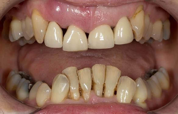
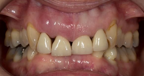
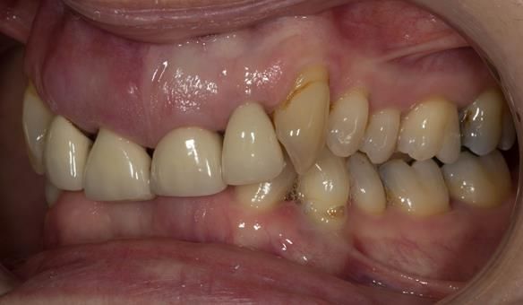
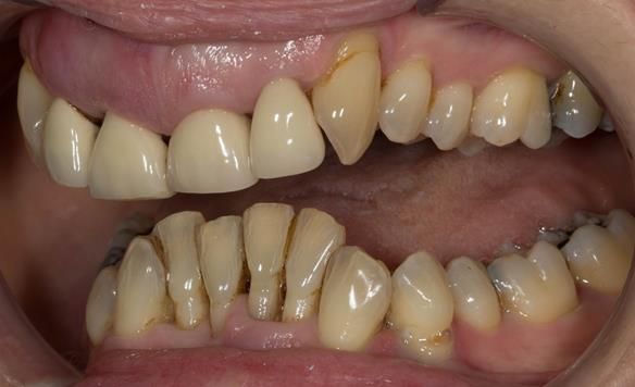
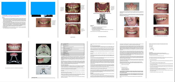
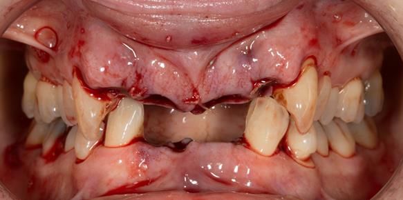
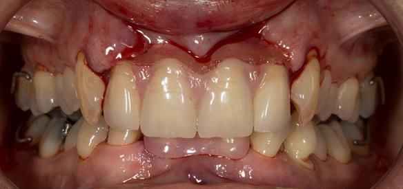
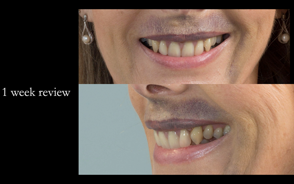
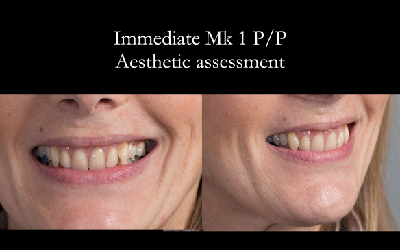
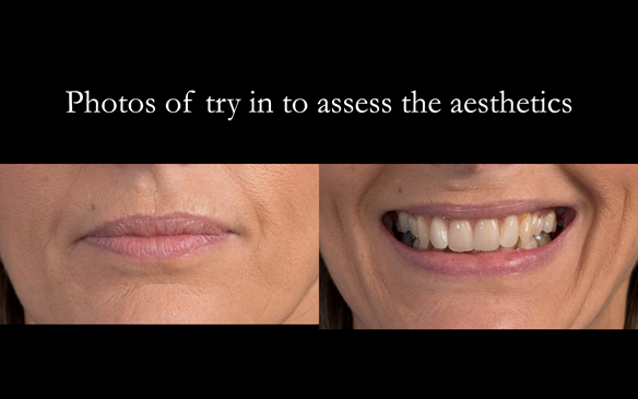
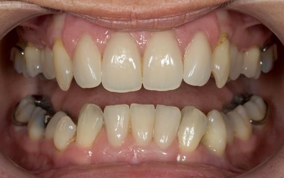
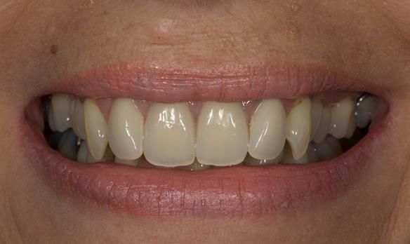
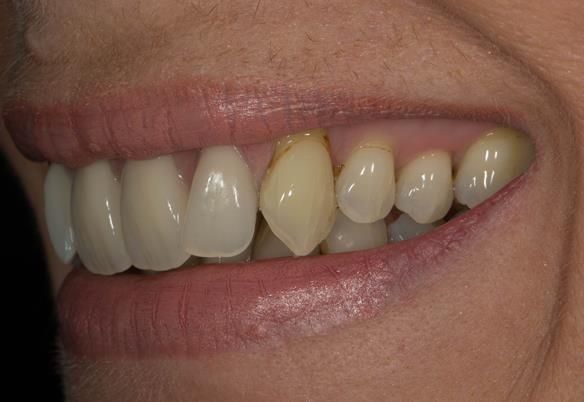
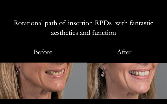
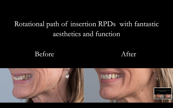
This has to be one of the most satisfying cases I have ever treated. The transformation in the patient's appearance and self confidence was incredible.

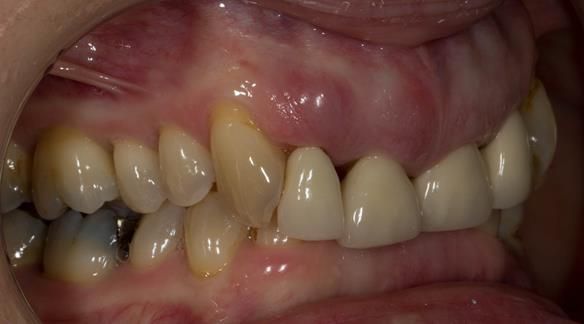
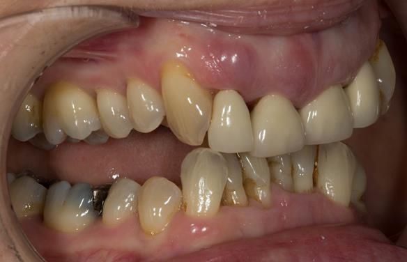
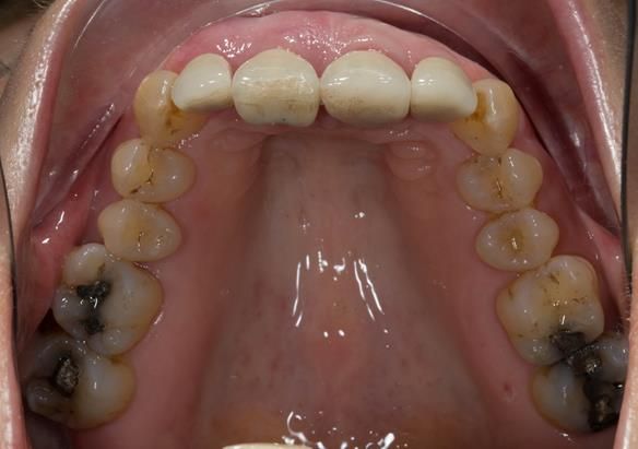
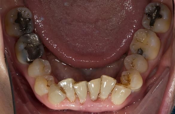
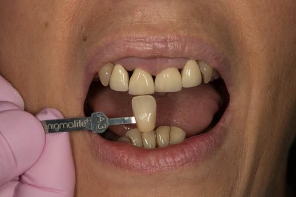
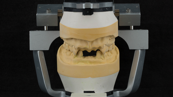
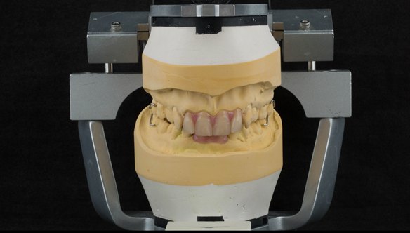
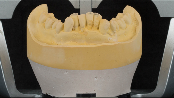
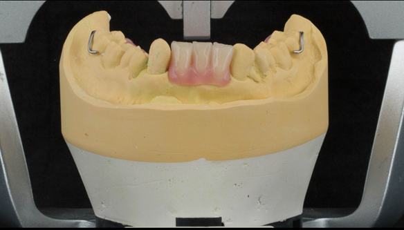
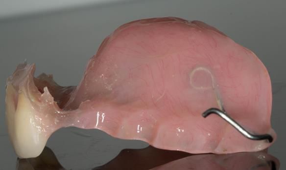
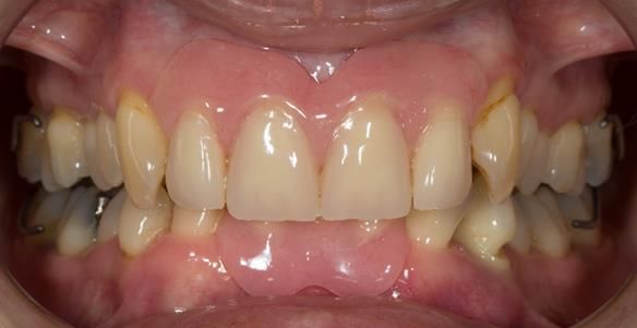
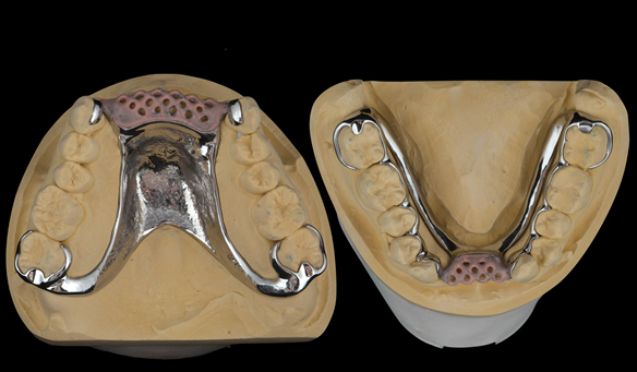
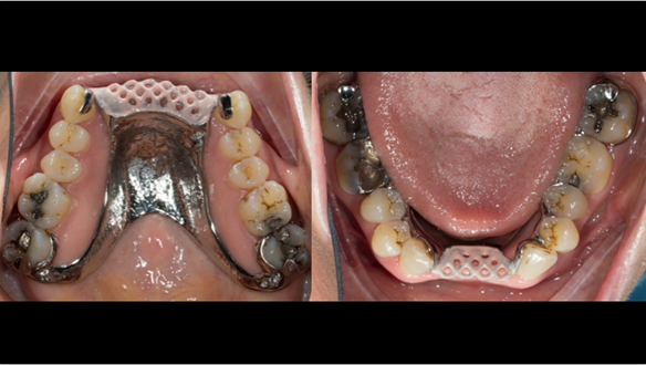
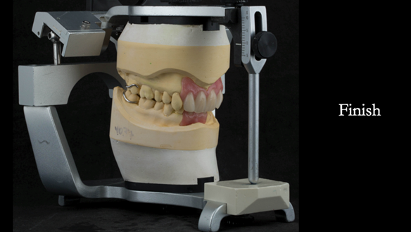
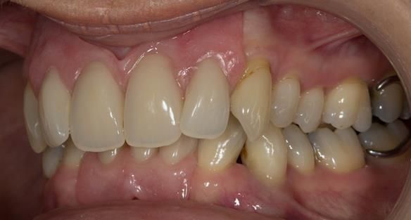
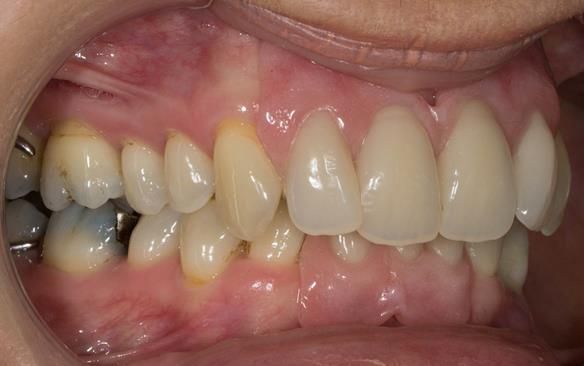
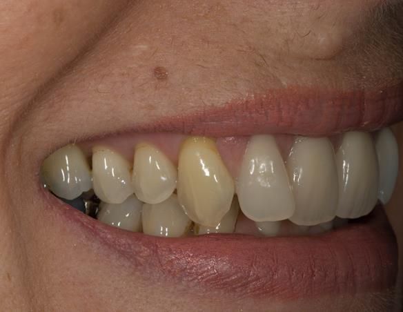
Transition from aesthetically failing, periodontally affected upper and lower incisors to definitive metal based upper and lower dentures. 57 year old woman.

Dental concerns

  • “Front teeth”
  • “Overbite”
  • “Gums”

Medical History: Fit and well

Dental wish list

  • “An improved smile”
  • “Healthy gums”

Diagnoses

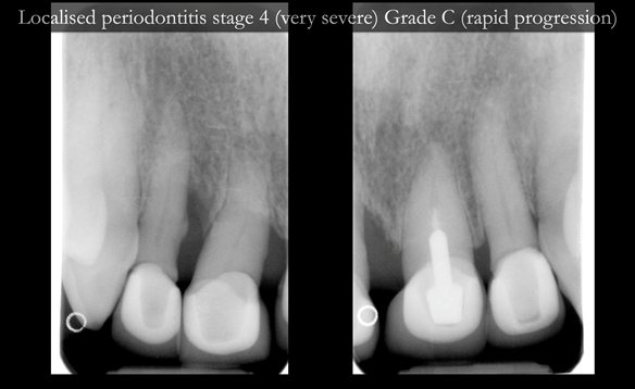
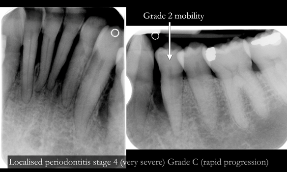
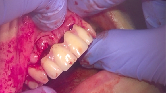
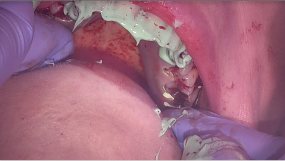
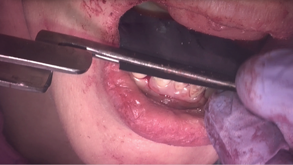
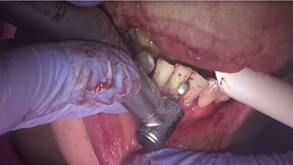
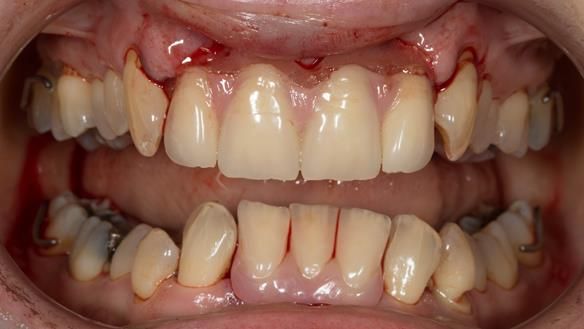
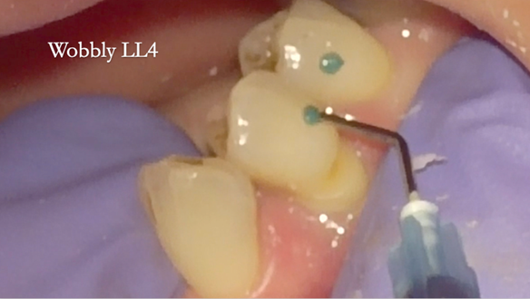
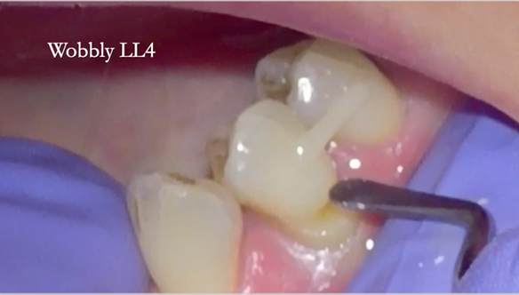
  • Localised periodontitis stage 4 (very severe) Grade C (rapid progression) - affecting the upper and lower incisors and LL4
  • Overeruption of the upper and lower anterior segments with traumatic overbite and aesthetic failure
  • Very high smile line
     

Treatment options

  • Do nothing
  • Periodontal management
  • Partial dentures
  • Bridges
  • Implant supported fixed teeth
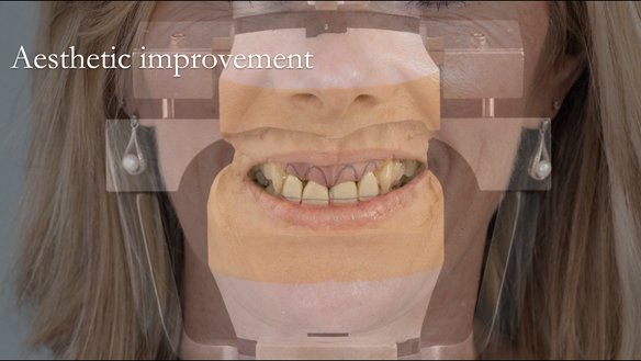
     

Treatment objective

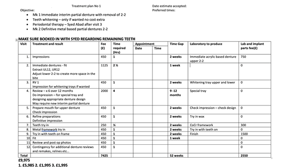
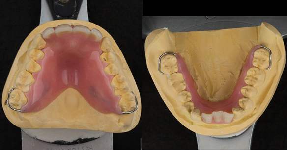
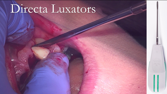
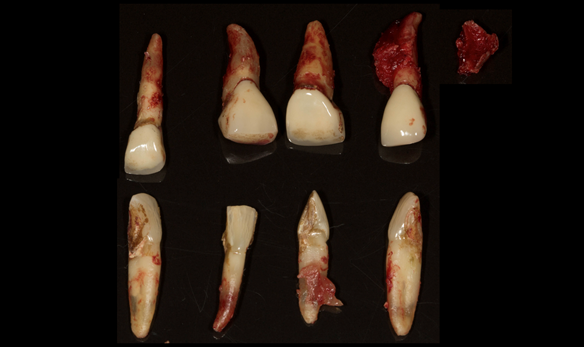
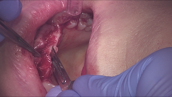
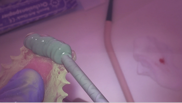
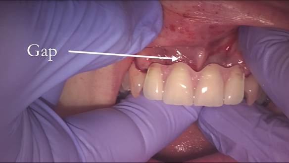
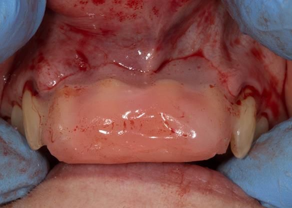
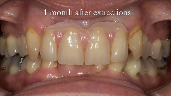
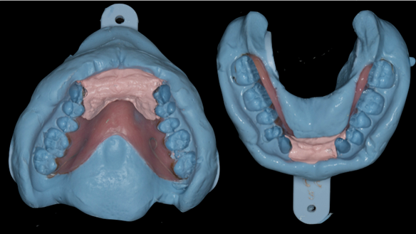
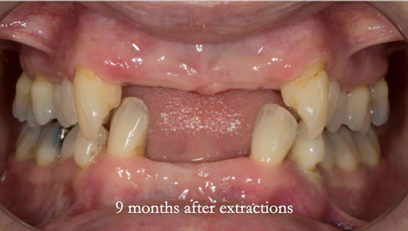
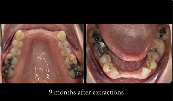
  • Mk 1 Immediate interim partial denture with removal of upper and lower 2-2
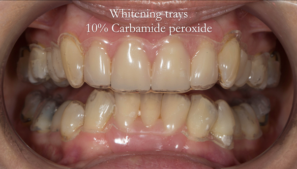
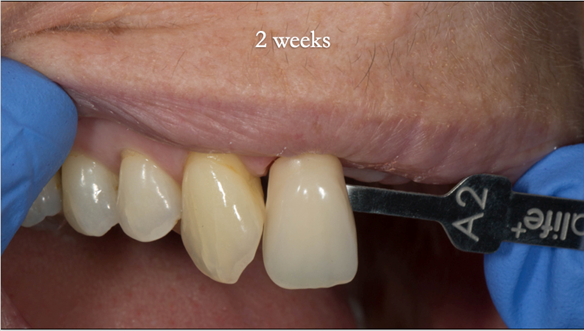
  • Teeth whitening – if wanted
  • Periodontal therapy
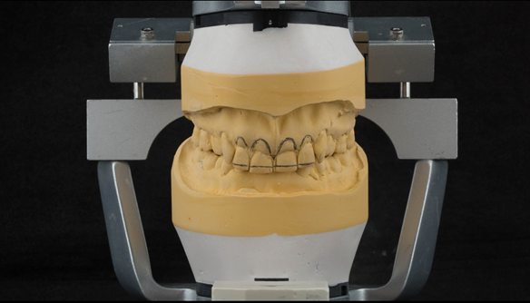
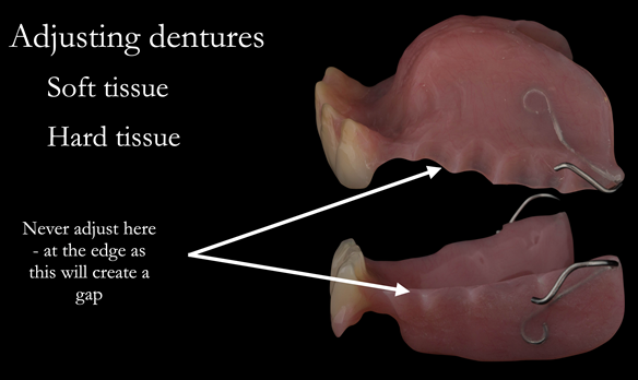
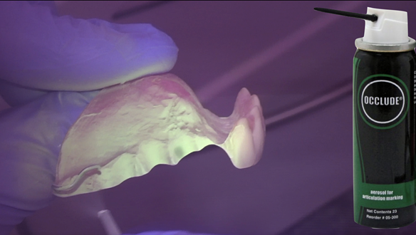
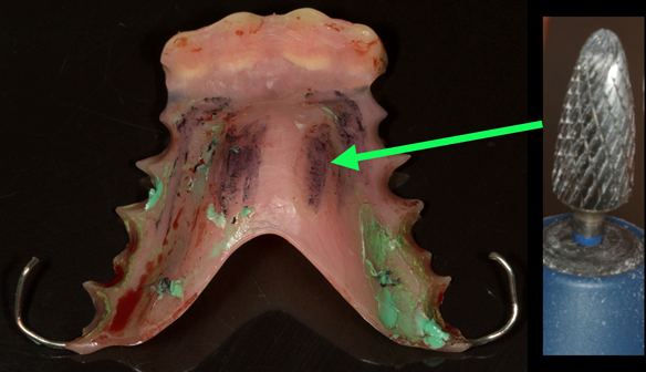
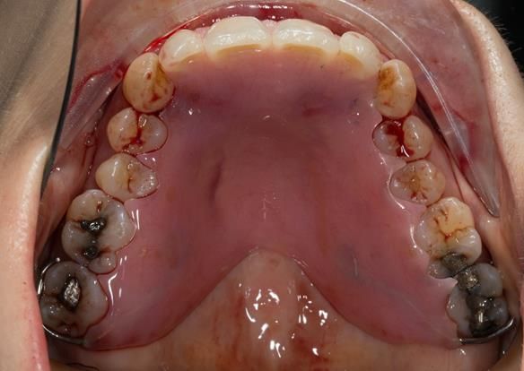
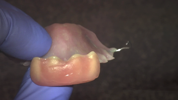
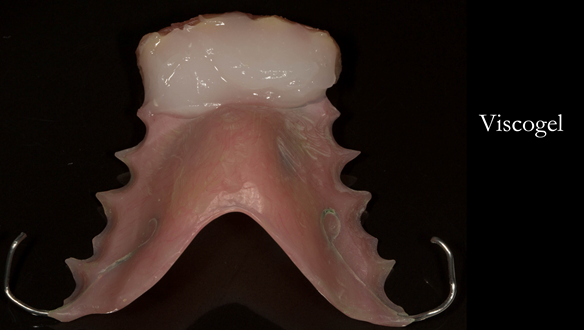
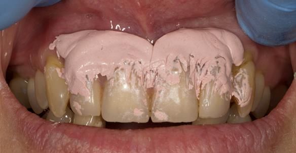
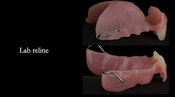
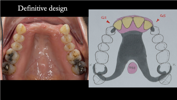
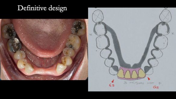
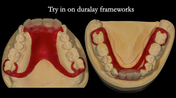
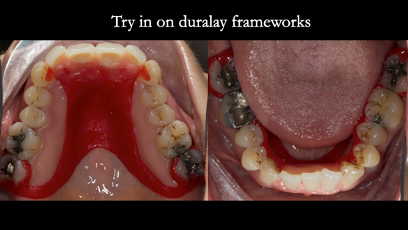
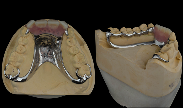
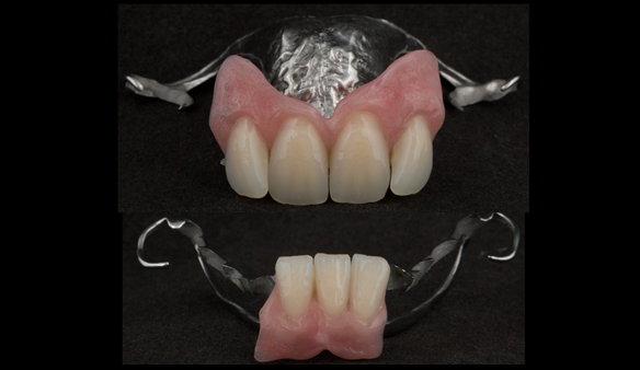
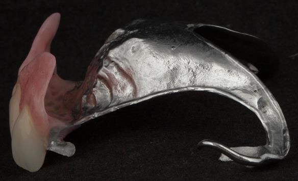
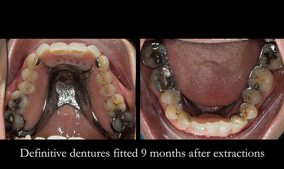
  • Mk 2 Definitive metal based partial dentures upper and lower 2-2

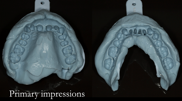
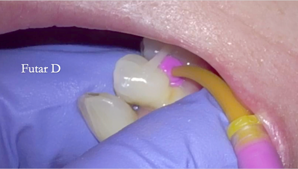
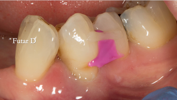
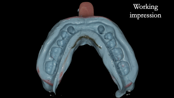
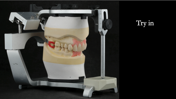
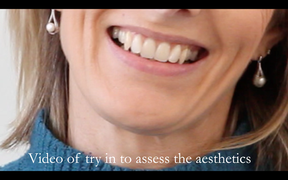
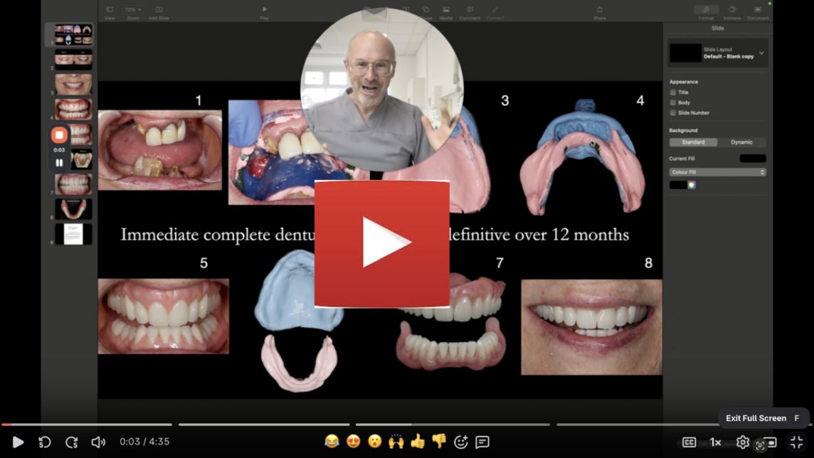
The clinical situation and treatment process is shown in detail below with photographs. I provided the clinical work. Rowan Garstang provided the technical work.

If you enjoy my Newsletters and you have friends, colleagues, dental students, dental technicians, clinical dental technicians and postgraduate dentists that you think will appreciate them, please feel free to share them. In addition, if you or your colleagues would like to receive my Newsletters and Denture Blog, please email me (education@finlaysutton.co.uk) and I will update the list.

Removable prosthodontic courses I run consisting of lectures and live clinical demonstrations:

These courses are aimed at general dentists, prosthodontists, clinical dental technicians and dental technicians. They are completely clinical, demonstrating how to provide dentures which have optimal function and superb aesthetics. I give live patient demonstrations along with lectures abundantly illustrated with step-by-step photographs and videos of all procedures. Delegates will take many “nuggets of gold”, being able to put the advice straight into practice with immediate improvement in professional satisfaction and patient outcomes.

Please see details for future courses given by Finlay over the next 2 years.

education@finlaysutton.co.uk

 Figure 1 Transition from aesthetically failing, periodontally affected upper and lower teeth to definitive metal based upper and lower dentures
Figure 1 Transition from aesthetically failing, periodontally affected upper and lower teeth to definitive metal based upper and lower dentures
Figure 2 Pre treatment very high smile line. Very difficult to produce good aesthetics with fixed prosthodontics
 Figure 3 Overeruption of the upper and lower anterior segments with traumatic overbite and aesthetic failure
Figure 3 Overeruption of the upper and lower anterior segments with traumatic overbite and aesthetic failure
 Figure 4 Overeruption of the upper and lower anterior segments with traumatic overbite and aesthetic failure
Figure 4 Overeruption of the upper and lower anterior segments with traumatic overbite and aesthetic failure
 Figure 5 Overeruption of the upper and lower anterior segments with traumatic overbite and aesthetic failure
Figure 5 Overeruption of the upper and lower anterior segments with traumatic overbite and aesthetic failure
 Figure 6 Overeruption of the upper and lower anterior segments with traumatic overbite and aesthetic failure
Figure 6 Overeruption of the upper and lower anterior segments with traumatic overbite and aesthetic failure
Figure 7 Overeruption of the upper and lower anterior segments with traumatic overbite and aesthetic failure
Figure 8 Overeruption of the upper and lower anterior segments with traumatic overbite and aesthetic failure
 Figure 9 upper arch crowns on upper anterior teeth preoperatively
Figure 9 upper arch crowns on upper anterior teeth preoperatively
Figure 10 lower arch preoperatively
Figure 11 Localised periodontitis stage 4 (very severe) Grade C (rapid progression) - affecting the upper incisors
Figure 12 Localised periodontitis stage 4 (very severe) Grade C (rapid progression) - affecting the lower incisors and LL4
Figure 13 treatment planning card containing sequenced treatment plan and quotation. This is how I plan all of my patients treatments
Figure 14 Treatment plan letter for the patient
Figure 15 Shade taking for immediate denture
Figure 16 Primary impression with Dentsply Blueprint
 Figure 17 Photograph of the patient's smile superimposed on the mounted working casts for the immediate dentures. Lines drawn on the cast for ideal replacement tooth positions.
Figure 17 Photograph of the patient's smile superimposed on the mounted working casts for the immediate dentures. Lines drawn on the cast for ideal replacement tooth positions.
Figure 18 Mounted working cast for Mk 1 immediate denture. Lines drawn on the cast for ideal replacement tooth positions.
Figure 19 Mounted working cast for Mk 1 immediate denture. Teeth removed from cast - minimal preparation of the cast to reduce adjustments at fit
Figure 20 Mounted working cast with Mk 1 immediate dentures. Figure 23 Finished upper and lower immediate dentures. Schottlander Enigmalife teeth
Figure 21 Mounted working lower cast for Mk 1 immediate denture. - showing overerupted teeth
Figure 22 Lower Mk 1 immediate denture with 3 incisors instead of 4 for improved appearance. Figure 23 Finished upper and lower immediate dentures. Schottlander Enigmalife teeth
Figure 23 Finished upper and lower immediate dentures. Schottlander Enigmalife teeth
Figure 24 Extraction of the upper and lower incisors using Directa Luxators
Figure 25 Extracted teeth
 Figure 26 When fitting I always adjust below area where the denture touches the tooth as in the photograph. This prevents a gap forming between the denture and the teeth when fitted as shown in figure 36
Figure 26 When fitting I always adjust below area where the denture touches the tooth as in the photograph. This prevents a gap forming between the denture and the teeth when fitted as shown in figure 36
Figure 27 Immediate denture not seating owing to thick soft tissue - incsial papilla
Figure 28 Thinning the incsial papilla and palatal tissues with number 15 blade
Figure 29 Extraction sites - sutures on the upper
Figure 30 Occlude spray is used to check the fit of the collets
 Figure 31 Light bodied silicone wash to check the soft tissue fitting surface.
Figure 31 Light bodied silicone wash to check the soft tissue fitting surface.
 Figure 32 Seating the immediate denture to check the fit with light bodied silicone and Occlude spray
Figure 32 Seating the immediate denture to check the fit with light bodied silicone and Occlude spray
Figure 33 The denture didn't seat properly on the palate. This was because of the primary impression shown in figure 16 didn't fully record the palate.
Figure 34 Once adjusted the denture seated correctly around the teeth as it does on the cast in figure 23
Figure 35 More alveolar tissue lost following extraction than anticipated. This space was filled in with Visco-gel as in the following slides
 Figure 36 Wax placed on labial surface of the teeth prior to Visco-gel reline. This prevents the viscogel from running into the interdental embrasures helping with quicker clean up
Figure 36 Wax placed on labial surface of the teeth prior to Visco-gel reline. This prevents the viscogel from running into the interdental embrasures helping with quicker clean up
 Figure 37 Temporary soft liner Visco-gel mixed ready to be placed in the mouth
Figure 37 Temporary soft liner Visco-gel mixed ready to be placed in the mouth
 Figure 38 Visco-gel temporary soft reline moulded in the mouth. The denture was seated fully with pressure on the palate
Figure 38 Visco-gel temporary soft reline moulded in the mouth. The denture was seated fully with pressure on the palate
 Figure 39 - Wax peeled off and Visco-gel can be trimmed and tidied with blade
Figure 39 - Wax peeled off and Visco-gel can be trimmed and tidied with blade
Figure 40 Occlusion checked
 Figure 41 Occlusion adjusted so that bite even on all of prosthetic teeth. In this case the bite was propped open on the the immediate dentures owing to a lack of space in ICP
Figure 41 Occlusion adjusted so that bite even on all of prosthetic teeth. In this case the bite was propped open on the the immediate dentures owing to a lack of space in ICP
 Figure 42 immediate denture fitted with patient in intercuspal position
Figure 42 immediate denture fitted with patient in intercuspal position
Figure 43 immediate denture fitted with teeth apart showing improved aesthetics with 3 lower incisor teeth
 Figure 44 1 week review - showing external bruising - this resolved in 2 weeks
Figure 44 1 week review - showing external bruising - this resolved in 2 weeks
Figure 45 Review 1 month after extractions. Gap has formed
 Figure 46 1 month after extractions - Immediate denture laboratory reline - part 1 with Zinc Oxide impression material
Figure 46 1 month after extractions - Immediate denture laboratory reline - part 1 with Zinc Oxide impression material
 Figure 47 1 month after extractions - Immediate denture laboratory reline - part 1 with Zinc Oxide impression material
Figure 47 1 month after extractions - Immediate denture laboratory reline - part 1 with Zinc Oxide impression material
 Figure 48 Immediate denture laboratory relines - part 2 alginate over impression in stock tray. Blueprint Dentsply.
Figure 48 Immediate denture laboratory relines - part 2 alginate over impression in stock tray. Blueprint Dentsply.
Figure 49 Immediate denture laboratory relined - this improves aesthetics and retention
 Figure 50 Relined immediate dentures 1 month after extractions
Figure 50 Relined immediate dentures 1 month after extractions
 Figure 51 Assessment of the aesthetics - immediate denture are great diagnostic dentures - improvements can be made in the definitive dentures
Figure 51 Assessment of the aesthetics - immediate denture are great diagnostic dentures - improvements can be made in the definitive dentures
 Figure 52 tooth-whitening trays made to fit over natural teeth and dentures
Figure 52 tooth-whitening trays made to fit over natural teeth and dentures
Figure 53 One shade lighter after two weeks of 10% Carbamide peroxide - nighttime whitening
Figure 54 definitive upper denture design
Figure 55 definitive lower denture design
 Figure 56 Check impression to assess preparation of the teeth for occlusal space for the rests
Figure 56 Check impression to assess preparation of the teeth for occlusal space for the rests
 Figure 57 Assessment of occlusal space of the check casts
Figure 57 Assessment of occlusal space of the check casts
 Figure 58 Working impression upper in custom tray with greenstick and alginate
Figure 58 Working impression upper in custom tray with greenstick and alginate
 Figure 59 Grade 2 mobile LR4 would move in the impression. To avoid this it was temporarily stabilised with a composite splint
Figure 59 Grade 2 mobile LR4 would move in the impression. To avoid this it was temporarily stabilised with a composite splint
Figure 60 Grade 2 mobile LR4 would move in the impression. To avoid this it was temporarily stabilised with a composite splint
 Figure 61 Grade 2 mobile LR4 would move in the impression. To avoid this it was temporarily stabilised with a composite splint and Futar D blocked out underneath to prevent impression tearing
Figure 61 Grade 2 mobile LR4 would move in the impression. To avoid this it was temporarily stabilised with a composite splint and Futar D blocked out underneath to prevent impression tearing
Figure 62 Grade 2 mobile LR4 would move in the impression. To avoid this it was temporarily stabilised with a composite splint and Futar D blocked out underneath to prevent impression tearing
 Figure 63 Working impression upper in custom tray with greenstick and alginate
Figure 63 Working impression upper in custom tray with greenstick and alginate
 Figure 64 Definitive casts mounted on Denar Mk 2 articulator. Teeth try in on pattern resin bases
Figure 64 Definitive casts mounted on Denar Mk 2 articulator. Teeth try in on pattern resin bases
 Figure 65 Teeth try in on pattern resin bases. These help to confirm the accuracy of the working casts and the amount of metal work which will be visible in the final dentures
Figure 65 Teeth try in on pattern resin bases. These help to confirm the accuracy of the working casts and the amount of metal work which will be visible in the final dentures
 Figure 66 Teeth try in on pattern resin bases in the mouth. These help to confirm the accuracy of the working casts and the amount of metal work which will be visible in the final dentures
Figure 66 Teeth try in on pattern resin bases in the mouth. These help to confirm the accuracy of the working casts and the amount of metal work which will be visible in the final dentures
 Figure 67 The patient assesses a video of the try in. This is a still shot of the video.
Figure 67 The patient assesses a video of the try in. This is a still shot of the video.
 Figure 68 The patient also assesses the aesthetics in photographs and in a mirror of the try in
Figure 68 The patient also assesses the aesthetics in photographs and in a mirror of the try in
Figure 69 cobalt chromium framework - Scandinavian design - keeping the denture components 3mm away from the gingival margin
 Figure 70 Cobalt chromium framework trial insertion, checking the fit onto the natural teeth
Figure 70 Cobalt chromium framework trial insertion, checking the fit onto the natural teeth
 Figure 71 Finished definitive partial dentures
Figure 71 Finished definitive partial dentures
 Figure 72 Finished definitive partial dentures. Scandinavian design - keeping the denture components 3mm away from the gingival margin
Figure 72 Finished definitive partial dentures. Scandinavian design - keeping the denture components 3mm away from the gingival margin
 Figure 73 Finished definitive partial dentures
Figure 73 Finished definitive partial dentures
 Figure 74 Finished upper definitive partial denture with knife edge flange - replacing the missing gum tissue. Anterior path of insertion to give retention
Figure 74 Finished upper definitive partial denture with knife edge flange - replacing the missing gum tissue. Anterior path of insertion to give retention
Figure 75 Ridges 9 months after extractions
Figure 76 Ridges 9 months after extractions
Figure 77 Definitive dentures fitted 9 months after extractions
 Figure 78 Definitive dentures fitted 9 months after extractions. Schottlander Enigmalife teeth
Figure 78 Definitive dentures fitted 9 months after extractions. Schottlander Enigmalife teeth
Figure 79 Definitive dentures fitted 9 months after extractions. Schottlander Enigmalife teeth
 Figure 80 Finished definitive partial dentures with knife edge flanges - replacing the missing gum tissue. Anterior path of insertion to give added retention
Figure 80 Finished definitive partial dentures with knife edge flanges - replacing the missing gum tissue. Anterior path of insertion to give added retention
 Figure 81 Finished definitive partial dentures with knife edge flanges - replacing the missing gum tissue. Anterior path of insertion to give added retention
Figure 81 Finished definitive partial dentures with knife edge flanges - replacing the missing gum tissue. Anterior path of insertion to give added retention
 Figure 82 Fitted cobalt chromium based partial dentures with Schottlander Enigmalife teeth
Figure 82 Fitted cobalt chromium based partial dentures with Schottlander Enigmalife teeth
 Figure 83 Fitted cobalt chromium based partial dentures with Schottlander Enigmalife teeth
Figure 83 Fitted cobalt chromium based partial dentures with Schottlander Enigmalife teeth
Figure 84 Fitted cobalt chromium based partial dentures with Schottlander Enigmalife teeth
 Figure 85 Before and after photos with cobalt chromium based partial dentures with Schottlander Enigmalife teeth
Figure 85 Before and after photos with cobalt chromium based partial dentures with Schottlander Enigmalife teeth
 Figure 86 Before and after photos with cobalt chromium based partial dentures with Schottlander Enigmalife teeth
Figure 86 Before and after photos with cobalt chromium based partial dentures with Schottlander Enigmalife teeth
 Figure 87 Before and after photos with cobalt chromium based partial dentures with Schottlander Enigmalife teeth
Figure 87 Before and after photos with cobalt chromium based partial dentures with Schottlander Enigmalife teeth
Rotational path of insertion RPDs with fantastic aesthetics FULL PROTOCOL
 I am giving a full day Masterclass on amazing dentures covering complete dentures, partial dentures and implant supported over dentures on Friday 3rd March 2023. Please contact Claus Krolak if you would like to come along.
I am giving a full day Masterclass on amazing dentures covering complete dentures, partial dentures and implant supported over dentures on Friday 3rd March 2023. Please contact Claus Krolak if you would like to come along.
Rotational path of insertion RPDs with fantastic aesthetics FULL PROTOCOL
 I am giving a two day Masterclass on amazing dentures covering complete dentures, partial dentures, immediate dentures and implant supported over dentures in Trondheim, Norway on Thursday 20th April and Friday 21st April 2023. Please contact Kai Arseth
I am giving a two day Masterclass on amazing dentures covering complete dentures, partial dentures, immediate dentures and implant supported over dentures in Trondheim, Norway on Thursday 20th April and Friday 21st April 2023. Please contact Kai Arseth
Rotational path of insertion RPDs with fantastic aesthetics FULL PROTOCOL
I am giving a two-day Masterclass on amazing dentures with a live patient demonstration covering complete dentures, partial dentures, immediate dentures and implant supported over dentures in Trondheim on Saturday 5th August and Sunday 6th April 2023. Ple
Rotational path of insertion RPDs with fantastic aesthetics FULL PROTOCOL
Finlay's Monthly Study Club Live Dates
A day with Finlay Sutton - designed for dentists and the team that hate making dentures
Finlay's Study Club Live 1 Managing a patient with overdentures on badly positioned implants
Massive suction on lower complete denture - full treatment walk through by Finlay Sutton

Finlay's Blog

Finlay's Monthly Study Club Live Dates

Each month I go through a case presentation from start to finish. Full of practical tips and tricks that can be taken straight into practice. I discuss problems and solutions I am having in clinical practice. It is free - click on the link to sign up to join. You can ask questions live in the chat box through out the meeting.

A day with Finlay Sutton - designed for dentists and the team that hate making dentures

Whether you love making dentures and want to do them better, have limited experience with them and want to learn more, or hate making dentures, this day is aimed at you. It will be a comprehensive guide to providing predictable dentures for your patients.

This course will be clinically useful if you work in the NHS, in private practice or in both. The day will be totally practical and clinical, with tips and tricks that can be put into practice on Monday morning.

The day will be peppered with nuggets of gold.

Finlay's Study Club Live 1 Managing a patient with overdentures on badly positioned implants - YouTube Video

This was stressful and challenging case I treated recently, both technically and managing the patient. I'll walk you through the process of providing complete dentures fitting over 6 very badly positioned dental implants. Please click on the link to watch the video of my talking through the case from start to finish.

Massive suction on lower complete denture - full treatment walk through by Finlay Sutton

Welcome to my Newsletter 62 video walk through, where I show the making and fitting of complete dentures plus extractions for Rafique. This issue provides a comprehensive overview of the entire protocol workflow.

prev
next