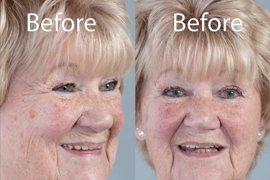
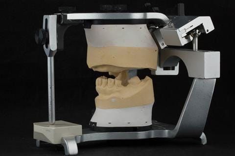
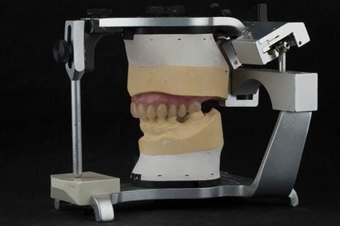
The 'gasket' or 'window' denture - provision of a maxillary cobalt chromium based partial denture
Diagnoses:
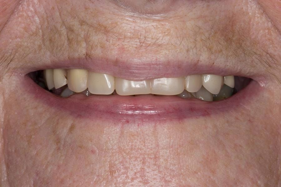
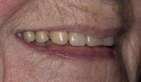
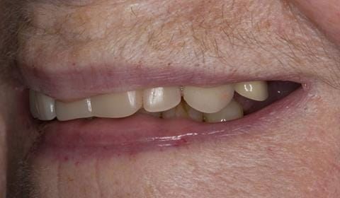
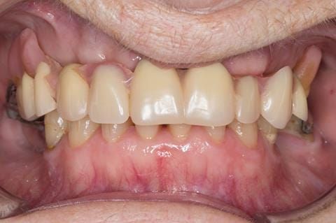
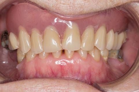
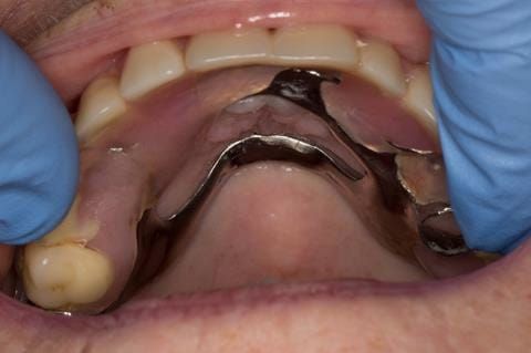
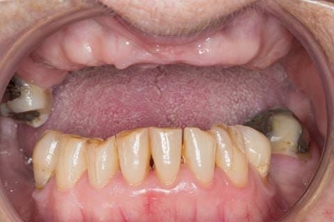
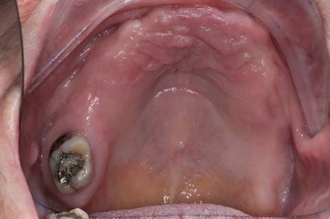
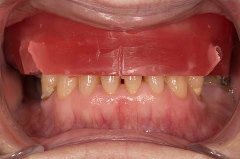
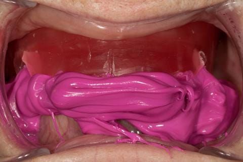
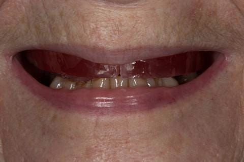
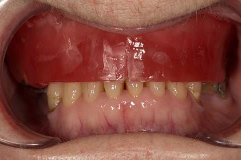
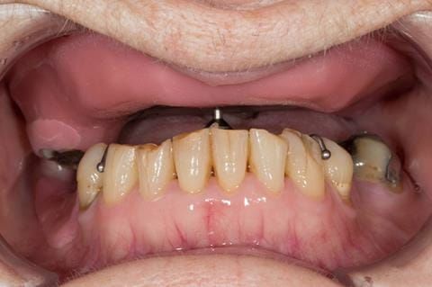
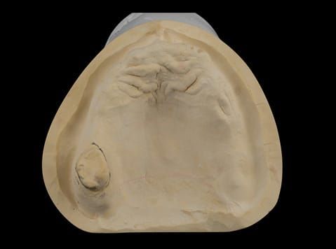
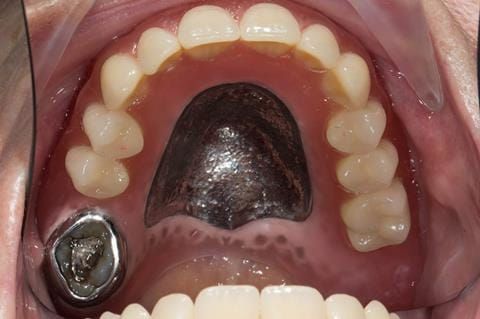
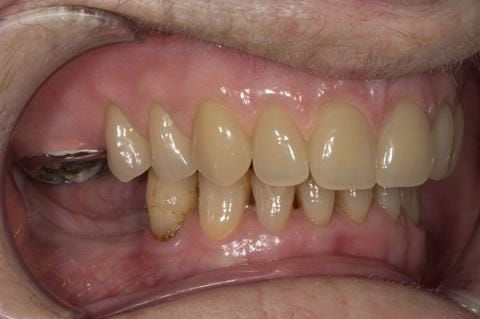
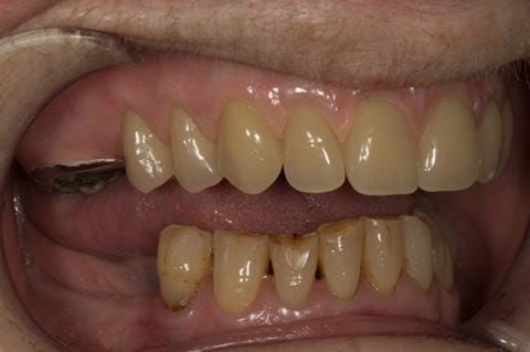
- Poorly fitting cobalt chromium based maxillary partial denture, which has been added to. This exhibited poor retention, stability and tissue fit (support). Unable to wear a new acrylic based denture.
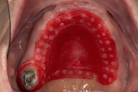
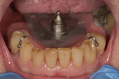
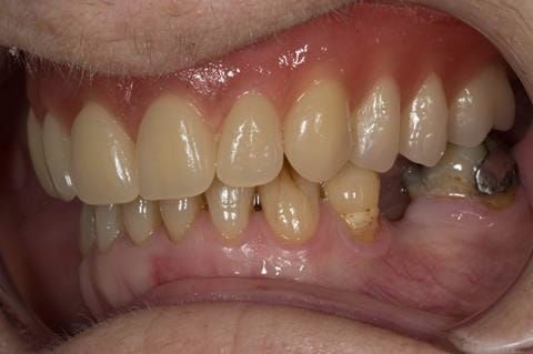
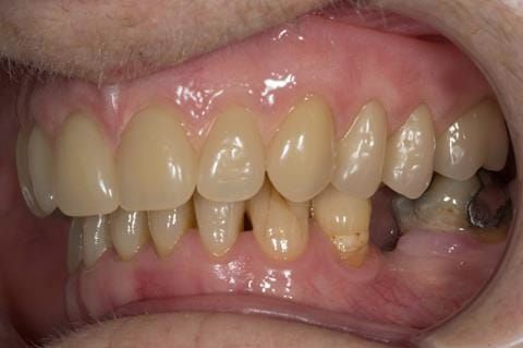
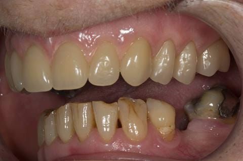
- UR7 - occlusal amalgam. 10- 20% alveolar bone loss. Healthy periodontium with reduced attachment level. No mobility.
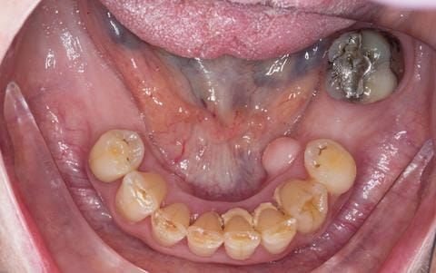
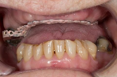
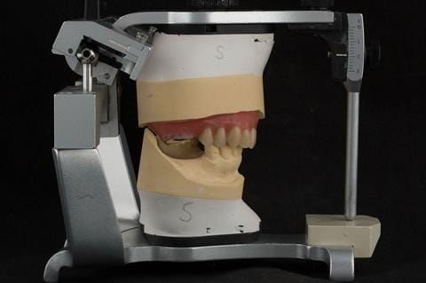
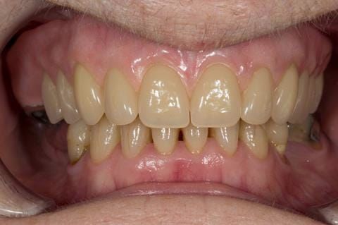
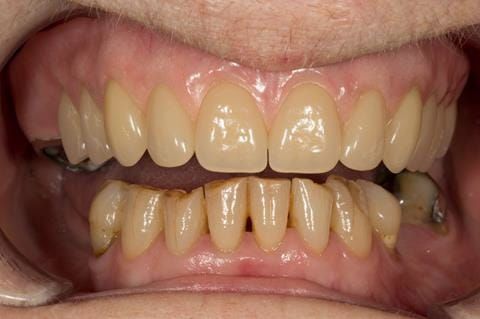
- Eight mandibular anterior teeth worn incisal edges from now extracted maxillary anterior crowns. Gingivitis - owing to inadequate oral hygiene.
- LL6 with large amalgam restoration - healthy periodontium.
- Bruxism.
Treatment options discussed with the patient to replace the missing maxillary teeth:
- Do nothing. The patient did not want this option.
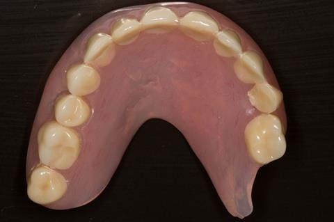
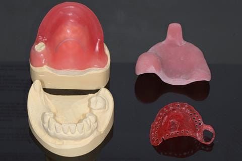
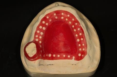
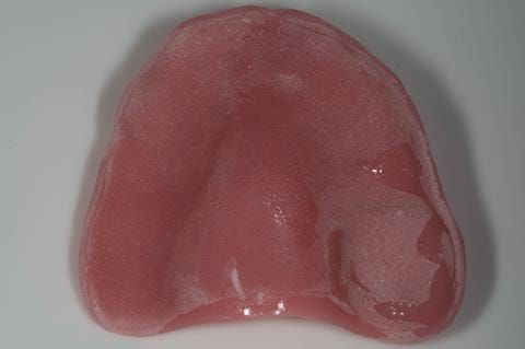
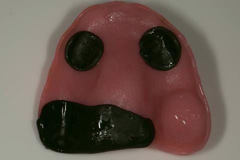
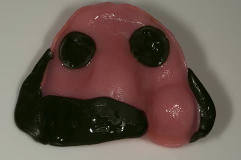
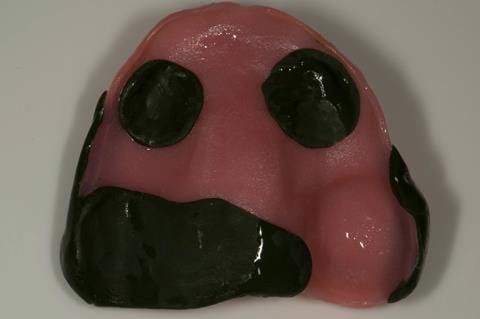
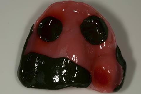
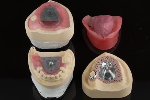
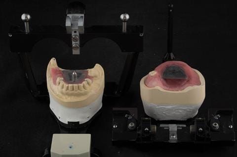
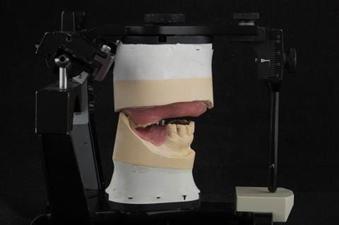
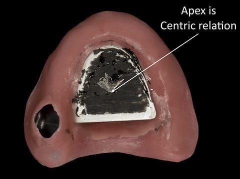
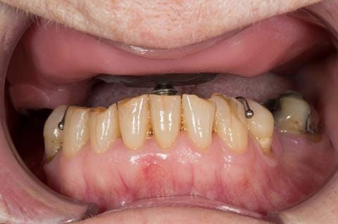
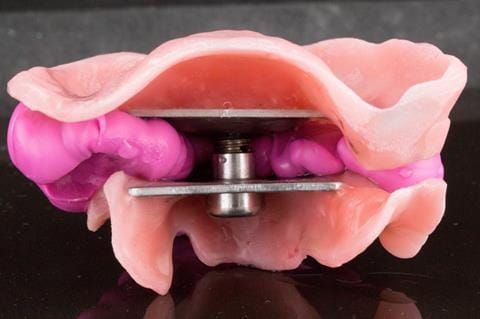
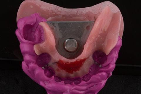
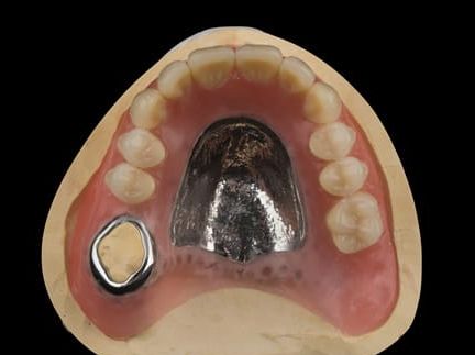
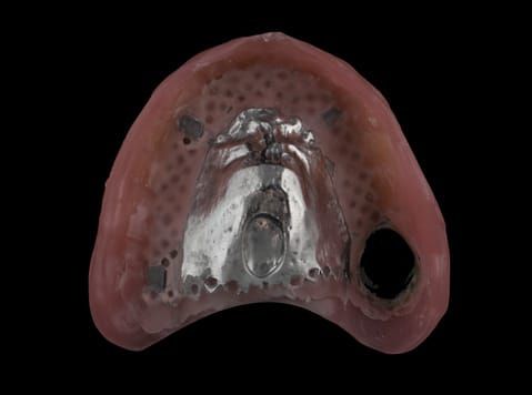
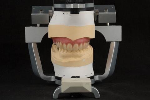
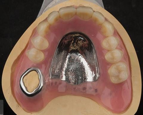
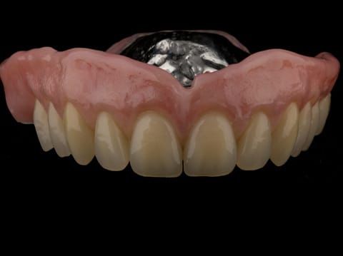
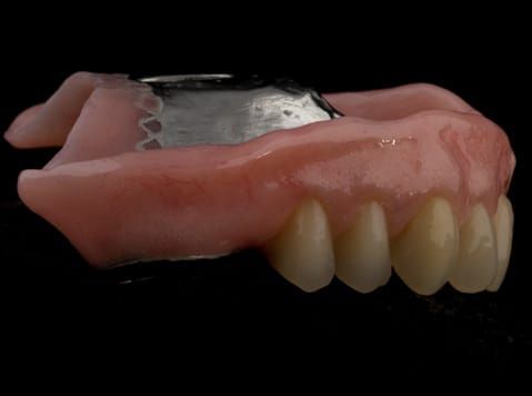
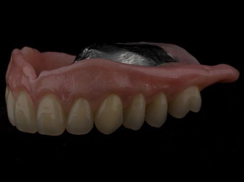
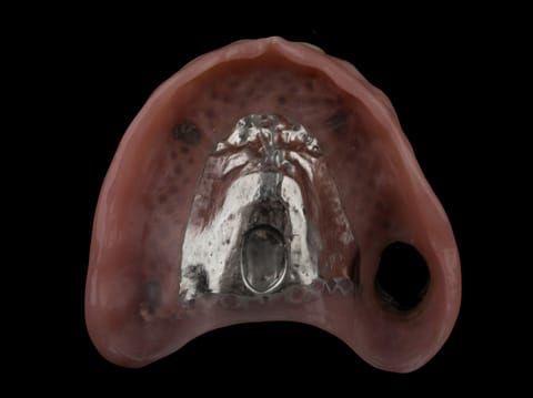
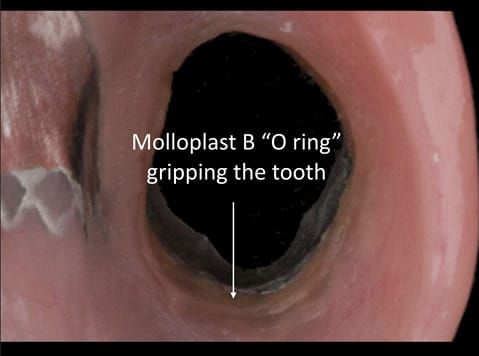
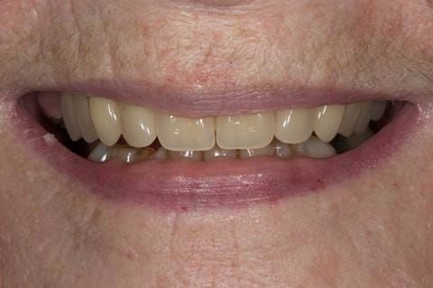
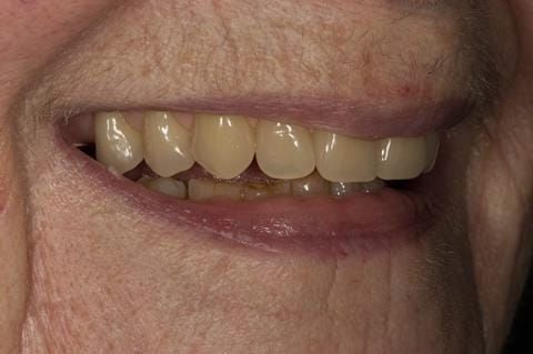
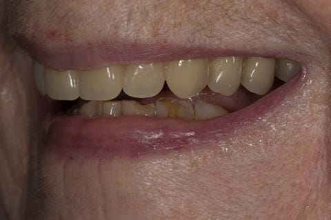
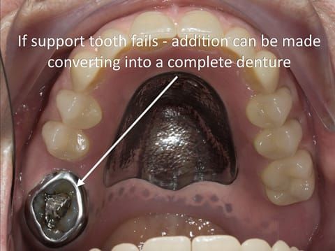
- Cobalt chromium reinforced gasket denture - using a Molloplast B "O" ring to retain and stabilise the denture. This was my professional preference as this was the least invasive and simplest solution to this dental problem. Should the UR7 require removal in the future - an artificial tooth could be added - resulting in a complete denture. The patient would have adapted to the denture fully by this stage and have good neuromuscular control of the prosthesis.
- Full upper denture fitted at the extraction of the remaining upper back tooth.
- Complete upper denture supported by dental implants.
- Fixed upper teeth supported by dental implants.
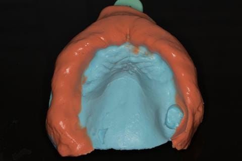
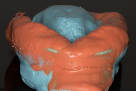
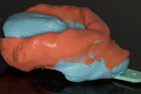
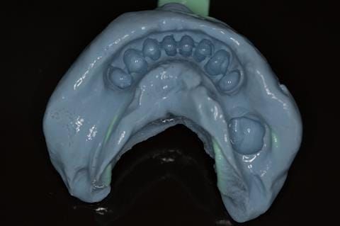
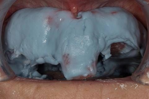
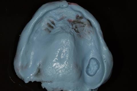
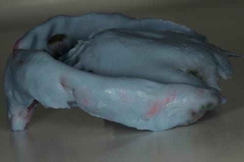
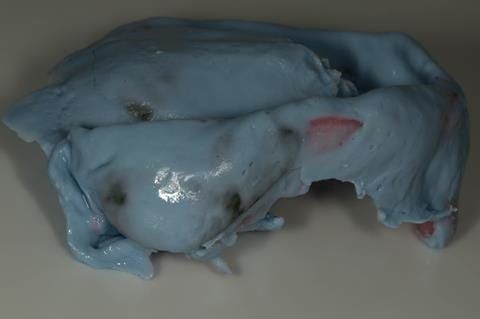
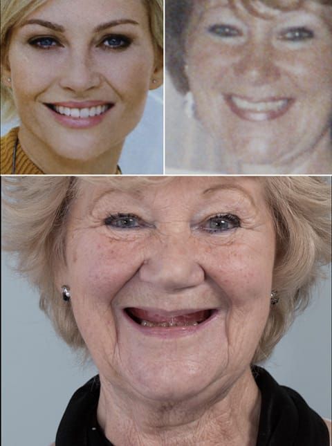
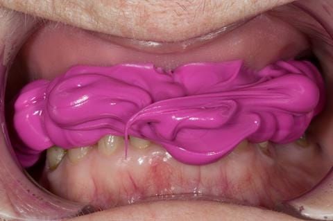
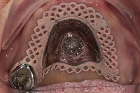
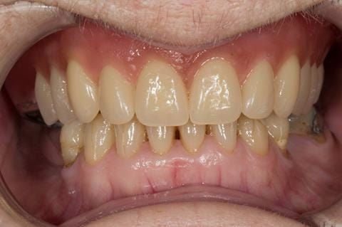
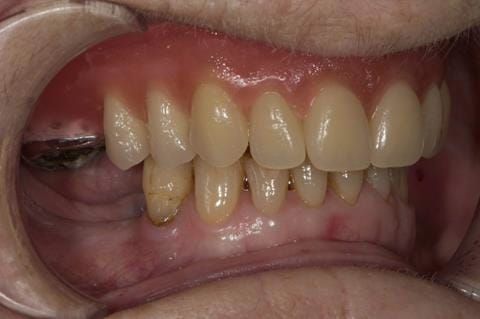
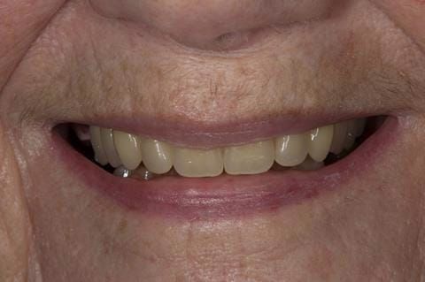
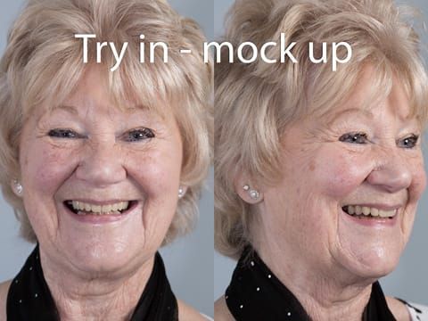
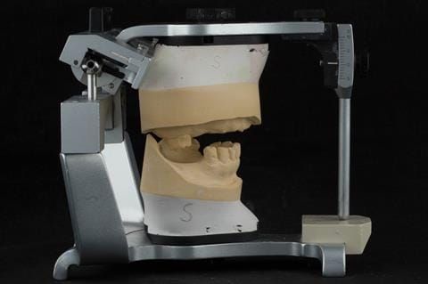
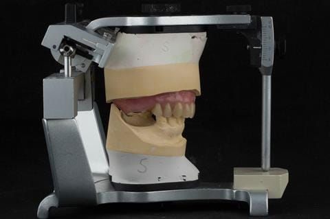
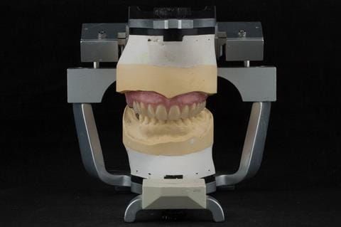
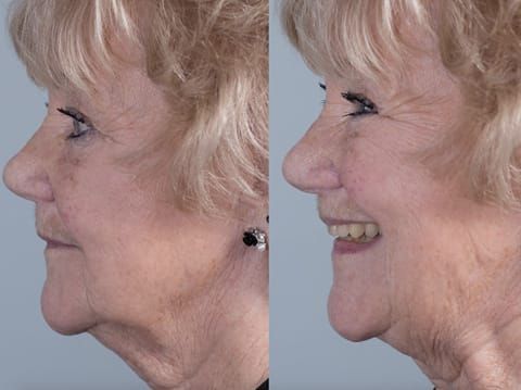
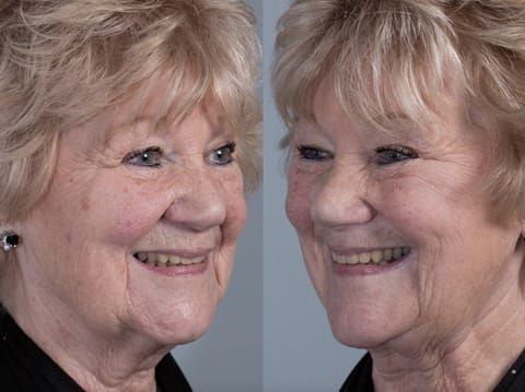
Following consultation and second discussion appointment the patient chose to have option 2 namely, a window denture - maxillary cobalt chromium based partial denture. The clinical situation and treatment process is shown in detail below with photographs. The patient was successfully rehabilitated with this and her quality of life considerably improved. The clinical work was provided by Finlay and the technical work by Rowan.