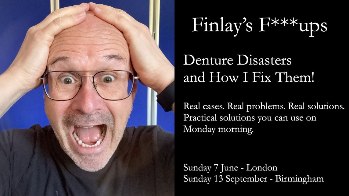
Covid-19 and my denture courses

** I plan on running my courses on complete dentures, partial dentures, immediate dentures and implant supported overdentures as soon as it is safe to do so. I will keep you up dated via Newsletters and my website. **

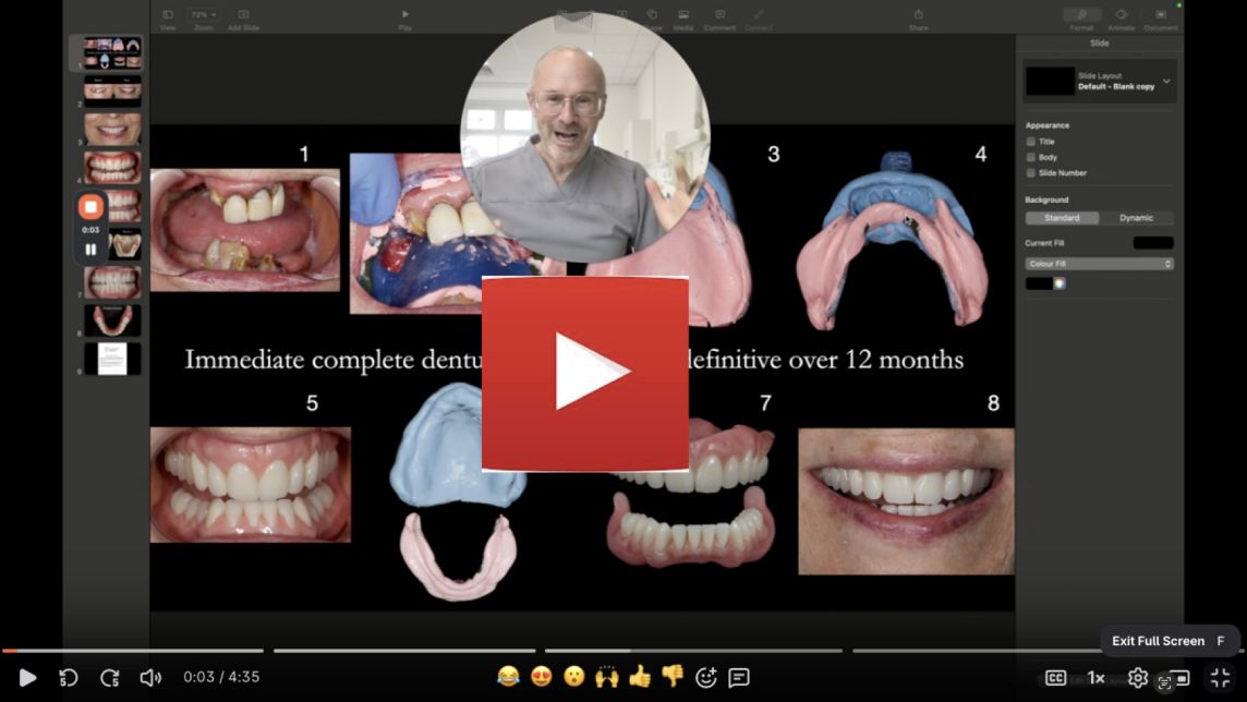
In the mean time I have been providing individual coaching, mentoring and treatment planning for removable prosthdontics via private recorded zoom meetings. They have been a great success with dentists all over the world taking part. The sessions are recorded so the mentee can refer back to the sessions. These can be one off or multiple sessions. Please click here for further information.

Finlay's Case Presentation

Welcome to my August 2020 Newsletter Case Presentation

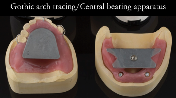
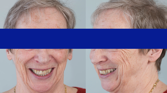
This newsletter describes in step by step detail the provision of an upper free end saddle partial denture in a patient with a high smile line and lower implant supported complete denture with suboptimal positioned dental implants.

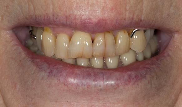
This 75 year old woman was referred to me from her general dental practiioner.

Dental History and Concerns

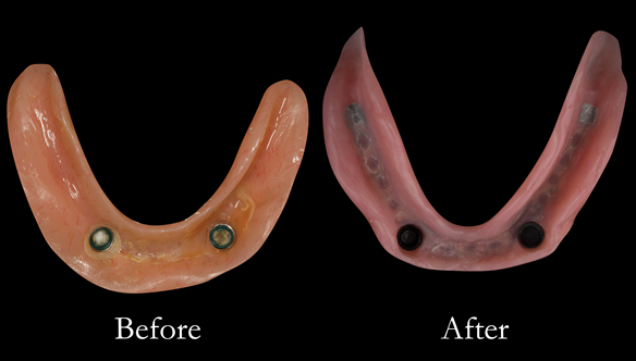
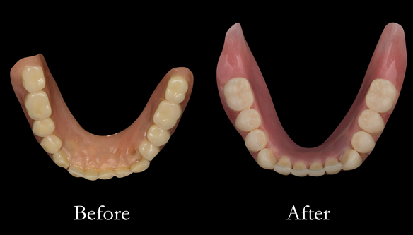
An implant supported lower complete lower denture had been provided 4 years previously in the UK. This "no longer fixes to its anchors and is worn". The upper partial denture was over 12 years old, having been provided in the USA. “Upper partial again worn and a little unreliable.”

Social History

Nil.

Dental wish list

“Good eating teeth – and ones/teeth that I am happy to smile with.”

Diagnoses

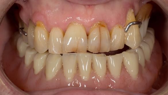
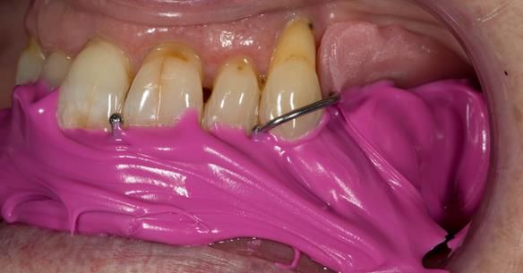
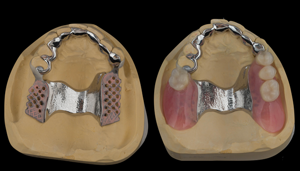
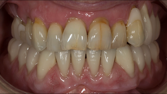
  1. The remaining natural upper 8 teeth had reduced but healthy periodontal attachment.
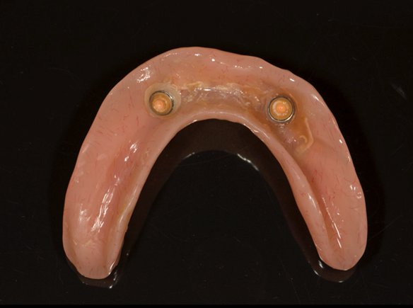
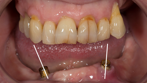
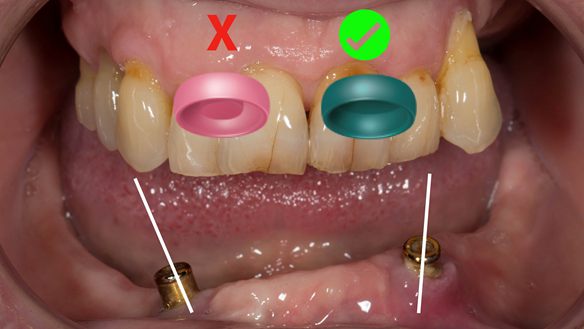
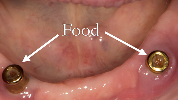
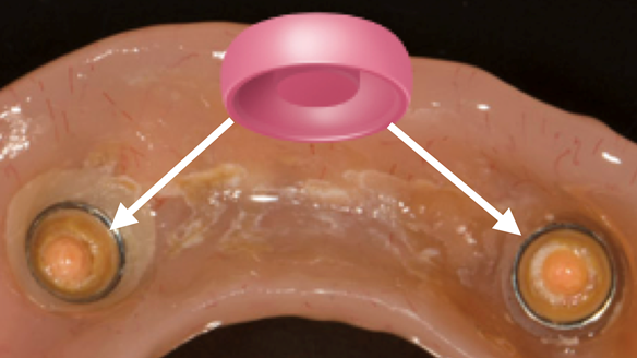
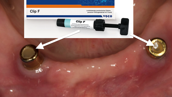
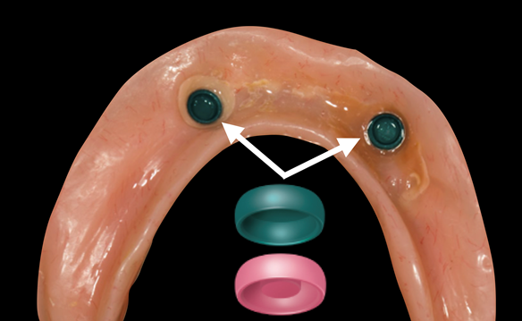
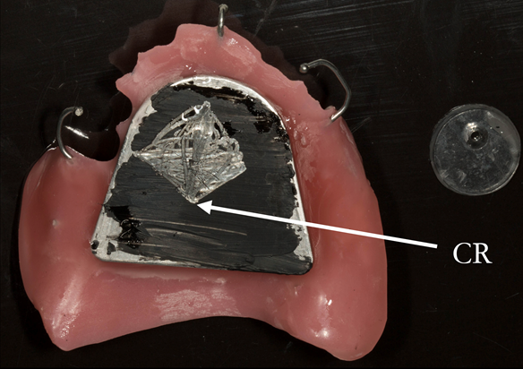
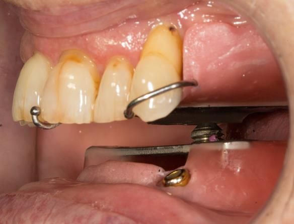
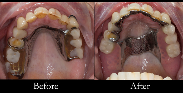
  2. The two dental implants (Astra) in the lower jaw had Locator attachments and appeared healthy. The implants positions were suboptimally positioned being over 30 degress divergant, resulting in poor fixation and increased wear of the Locator components.
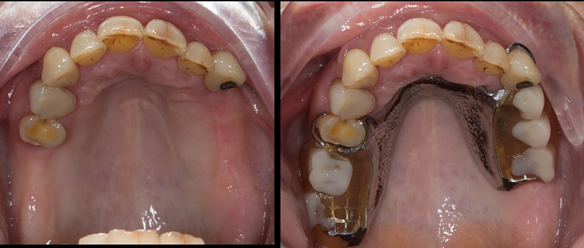
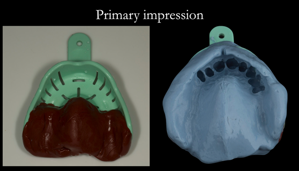
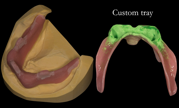
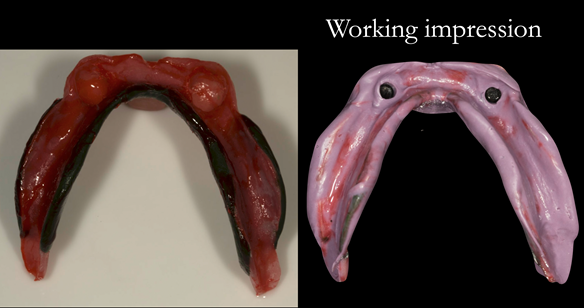
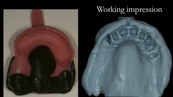
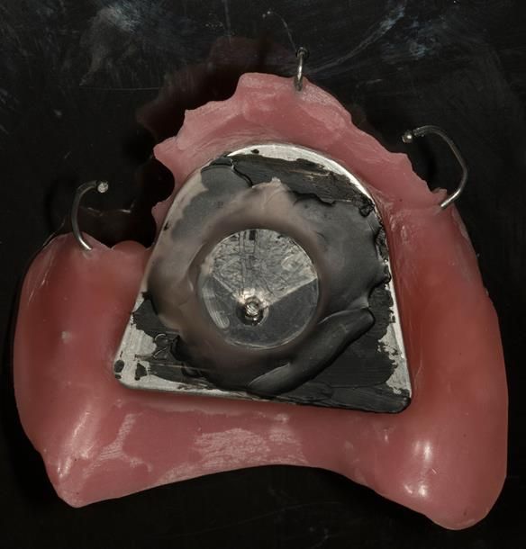
  3. The upper and lower dentures exhibited suboptimal extension of the flanges and saddles. They had poor retention, support and stability.
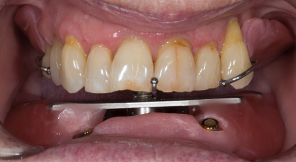
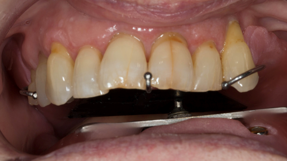
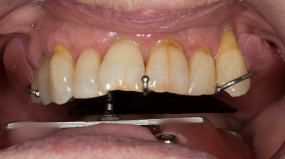
  4. The patient had a high smile line showing the cobablt chromium clasps of the UR4 and UL3 during social interaction.

Apart from providing no treatment we discussed two solutions namely; fixed implant supported teeth versus optimally designed and made removable dentures. The patient chose to have removable partial dentures.

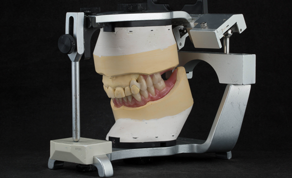
The clinical situation and treatment process is shown in detail below with photographs. I provided the clinical work and Rowan Garstang provided the technical work.

If you enjoy my Newsletters and you have friends, colleagues, dental students, dental technicians, clinical dental technicians and postgraduate dentists that you think will appreciate them, please feel free to share them. In addition, if your colleagues would like to receive our Newsletters please email me and I will add them to my email list.

Figure 1 Pre - treatment. High smile line showing metal clasp.
Figure 2 Pre - treatment. High smile line showing metal clasp. Upper partial denture and lower complete denture.
Figure 3 Pre - treatment. High smile line showing metal clasp. Upper partial denture and lower implant supported complete denture.
Figure 4 The remaining natural upper 8 teeth reduced but healthy periodontal attachment. Upper denture with suboptimal extensions.
Figure 5 Pre-treatment lower implant supported complete denture. Under-extended in retromolar area and overextended labially, indicating a lack of border moulding during impression taking.
Figure 6 Divergent implants - over 30 degrees
Figure 7 Green locator attachments (without male insert) work better on divergent dental implants
Figure 8 Food (particularly seeds) get pushed into the abutment centre resulting in the denture attachment not seating
Figure 9 This results in deformation damage to the attachments
Figure 10 A superb tip Prof Frauke Muller taught me. A temporary filling material placed into the top of the abutment prevents food from collecting
Figure 11 The attachment inserts were then changed from pink to green to allow the denture to seat
Figure 12 treatment planning card containing sequenced treatment plan and quotation. This is how I plan all of my patients treatments
Figure 13 Lower primary impression with two viscosities using Zhermack Tropcalgin (base) and Neocolloid (Syringe). This allows full extension to record the sulcui
Figure 14 Upper primary impression - 2 stages compound first to record the free end saddles and alginate medium wash
Figure 15 Custom tray for lower working impression. This is adjusted so that the impression pick ups for the locators do not touch the inside of the tray when this is seated in the mouth - ensures correct seating of the tray
Figure 16 Lower working impression. Border moulded in greenstick followed by Impregum impression with full border moulding
Figure 17 Upper working impression. Border moulded in greenstick followed by alginate impression with full border moulding
Figure 18 Registration visit with central bearing apparatus (gothic arch tracing) for CR recording
Figure 19 Registration visit with central bearing apparatus (gothic arch tracing) for CR recording
Figure 20 Registration visit with central bearing apparatus (gothic arch tracing) for CR recording moving to the patient's left
Figure 21 Registration visit with central bearing apparatus (gothic arch tracing) for CR recording moving to the patient's right
Figure 22 Inter-maxillary registration with central bearing apparatus. Maxillary plate with china graph pencil marking with arrow head scribed showing precise CR
Figure 23 Inter-maxillary registration with central bearing apparatus. Maxillary plate with plastic disc with countersunk hole placed over CR arrow head. This allows the mandibular pin to fit into the hole to enable accurate recording of this inter-maxill
Figure 24 Inter-maxillary registration with central bearing apparatus in the mouth with the maxillary and mandibular parts "locked" together in CR by the pin in the hole
Figure 25 Inter-maxillary registration with central bearing apparatus in the mouth with the maxillary and mandibular parts registered together with futar D bite registration material
Figure 26 Mounted casts with finished dentures on the articulator. Schottlander Enigmalife denture teeth and Dental D clasps
Figure 27 Before and after changes in shape of the denture fitting surfaces. Optimal extensions from border moulding. New denture metal reinforced too
Figure 28 Before and after changes in shape of the denture fitting surfaces. Optimal extensions from border moulding. New denture metal reinforced too
Figure 29 Upper denture cobalt chromium base and finished denture. Optimal extension of saddles and palatal strap for maximum support. Dental bar gives additional support and should teeth be lost in the future this can be added to
Figure 30 Finished upper denture. Optimal extension of saddles and palatal strap for maximum support. Dental bar gives additional support and should teeth be lost in the future this can be added to
Figure 31 Finished denture mimicking the natural teeth. Schottlander Enigmalife denture teeth and Dental D clasps
Figure 32 Finished denture mimicking the natural teeth. Schottlander Enigmalife denture teeth and Dental D clasps
Figure 33 Finished denture mimicking the natural teeth. Schottlander Enigmalife denture teeth and Dental D clasps

Reference material

Full access PDF to my published scientific papers which explain my philosophy and clinical techniques. Please click on the link below and scoll down this page to find lots of useful clinical techniques, reference material and previous lectures:

https://www.finlaysutton.co.uk/speaking

Massive suction on lower complete denture - full treatment walk through by Finlay Sutton
One to One Removable Prosthodontics Denture Mentorship with Finlay
Finlay designs a Kennedy class 1 upper metal based partial denture for Sue in real time.
Finlay designs a Kennedy class 2 (1) metal based partial denture for Sylvia - who has had MRONJ - VIDEO ON YouTube

Finlay's Blog

Massive suction on lower complete denture - full treatment walk through by Finlay Sutton

Welcome to my Newsletter 62 video walk through, where I show the making and fitting of complete dentures plus extractions for Rafique. This issue provides a comprehensive overview of the entire protocol workflow.

One to One Removable Prosthodontics Denture Mentorship with me, Finlay

Hi there! I'm here to help you achieve the best results with removable pros. I can assist with complete dentures, interesting partials, and even implants over dentures. Let's work together to reduce reviews, make patients happier, and provide better outcomes. If you need mentorship, coaching, or treatment planning, reach out to me at education@finlaysutton.com.

The cost for mentorship is £450 per hour, plus VAT.

Finlay designs a Kennedy class 1 upper metal based partial denture for Sue in real time

Finlay designs a Kennedy class 1 upper metal based partial denture for Sue in real time.

This is how I design RPDs for all of my cases. YouTube Video.

Finlay designs a Kennedy class 2 (1) metal based partial denture for Sylvia - who has had MRONJ - VIDEO ON YouTube

Finlay designs a Kennedy class 2 (1) metal based partial denture for Sylvia - who has had MRONJ - VIDEO ON YouTube

prev
next